Purpose
Background and importance. ILNEB syndrome (interstitial lung disease, nephrotic syndrome, and epidermolysis bullosa) is a rare autosomal-recessive disorder caused by biallelic variants in ITGA3, which encodes the alpha-3 subunit of the alpha-3/beta-1 (α3β1) integrin. The pulmonary phenotype is severe and progressive, yet its molecular architecture is poorly resolved because affected patients are exceptionally rare and no ITGA3-loss cohort exists. Standard idiopathic pulmonary fibrosis (IPF) genetic testing does not capture this mechanism, and the histologic pattern is frequently misassigned to alveolar usual interstitial pneumonia (UIP).
Case summary. We report a 31-year-old male with compound-heterozygous ITGA3 variants, a 2014 living-related kidney transplant for ITGA3-associated renal fibrosis, chronic post-transplant immunosuppression, and severe mixed restrictive and obstructive lung disease requiring continuous supplemental oxygen. He underwent lung-transplant evaluation in July 2025. Two lung biopsies collected five years apart (2017 and 2022) and a 2025 blood draw enabled a longitudinal, seven-modality analysis.
Molecular findings. The genotype comprises one null allele (c.2070+1G>A, canonical splice-donor disruption predicted to trigger nonsense-mediated decay) and one hypomorphic missense allele (c.1973C>G, p.Thr658Arg), both clustering in the Calf-1 domain and classified as Pathogenic / Likely Pathogenic. A parallel, genotype-independent epigenetic axis was identified: MUC5B and TERT promoter hypermethylation despite a wild-type MUC5B risk allele (rs35705950 G/G). Integrated across genomics, plasma proteomics, single-cell and long-read transcriptomics, and paired spatial modalities, the data support an epithelial-skewed, regulatory (non-inflammatory) peripheral profile with epithelial-injury markers (KRT19, SPP1) dominant. The spatial comparison shows a major immune reorganization between 2017 and 2022, with an approximately eight-fold tissue T-cell expansion, airway basal-epithelial collapse, and stable-to-decreasing myofibroblast burden, together reframing the disease as small-airway / constrictive bronchiolitis (bronchiolitis obliterans syndrome, BOS-like) rather than alveolar UIP.
Clinical implications. The BOS reframe and the second epigenetic axis redirect therapeutic reasoning away from blunt pan-TGF-beta ligand blockade and toward phenotype-matched, cGVHD-BOS-relevant agents, IL-6 blockade, and DNMT-directed epigenetic candidates, all constrained by patient-specific pharmacogenomics. This case demonstrates how integrative causal multi-omics can extract clinically actionable structure from a single rare-disease patient.
Introduction
Integrin α3β1 is an epithelial cell-adhesion receptor that anchors basal and airway epithelial cells to the basement membrane and participates in epithelial repair, adhesion, and mechanotransduction. Biallelic loss-of-function (LOF) variants in ITGA3 cause ILNEB syndrome, a rare autosomal-recessive condition combining interstitial lung disease, nephrotic-range renal disease, and skin fragility. Because the receptor is required for epithelial integrity, ITGA3 loss is expected to produce an epithelial-primary form of lung disease that is mechanistically distinct from common IPF.
Two features complicate clinical reasoning in these patients. First, the canonical IPF genetic risk architecture (for example the MUC5B promoter variant rs35705950 and telomere-maintenance genes) does not explain ITGA3-driven disease, so standard testing under-informs both diagnosis and prognosis. Second, the pulmonary histology can resemble either alveolar UIP or a small-airway, constrictive bronchiolitis (BOS-like) pattern, and the two framings imply different therapeutic priorities.
The core gap this study addresses is that no ITGA3-LOF cohort exists, so mechanism must be inferred at the level of a single patient. We approach this by mapping seven molecular modalities, spanning two timepoints five years apart, onto a literature-derived causal directed acyclic graph (DAG) and testing which mechanistic edges the patient’s own data confirm. The objective is not population-level generalization but a rigorously traced, patient-specific mechanistic model that can guide diagnosis, prognosis, and repurposing hypotheses.
Case Presentation
Demographics and history. A 31-year-old male carries compound-heterozygous ITGA3 variants causing ILNEB syndrome. He has a history of renal fibrosis and received a living-related donor kidney transplant in 2014 without complication or subsequent rejection, and he is maintained on chronic post-transplant immunosuppression (tacrolimus and mycophenolate mofetil).
Presentation. He presents with severe pulmonary disease characterized by extensive fibrosis, obstructive and constrictive bronchiolitis, and chronic respiratory failure requiring continuous supplemental oxygen. Progressive symptoms led to lung-transplant evaluation in July 2025.
Pulmonary function (October 2025).
| Parameter | Value | % Predicted | Interpretation |
|---|---|---|---|
| FVC | 1.64 L | 37.1% | Severe restriction |
| FEV1 | 0.92 L | 24.8% | Severe obstruction |
| DLCO | 12.54 | 42.5% | Moderate gas-transfer impairment |
These values indicate a severe mixed restrictive and obstructive limitation.
Imaging and pathology. Retrospective high-resolution chest CT from 2016 shows combined interstitial fibrosis and bronchiolitis obliterans, with significant worsening since 2019 but minimal change since 2024. Parametric response mapping (February 2026) demonstrates consolidative opacities and air trapping (functional small-airway disease, fSAD approximately 8%). Upper-lobe wedge-resection pathology demonstrates a cGVHD BOS-like phenotype: bronchiolitis obliterans with lymphocytic bronchiolitis and associated lymphoid aggregates. These small-airway changes progressed from the 2017 wedge resection, directly corroborating the 2017-to-2022 spatial progression described below. Notably, the patient did not receive an allogeneic hematopoietic stem-cell transplant, so the BOS-like designation is a morphologic pattern rather than a shared etiology.
Regulatory T-cell status. T-regulatory-cell-specific demethylated region (TSDR) analysis of peripheral blood mononuclear cells (PBMCs) demonstrated reduced regulatory T cells, providing an orthogonal blood-based signal of current Treg deficiency at a single timepoint.
Current medications (abbreviated). Tacrolimus and mycophenolic acid (transplant-mandated immunosuppression); nerandomilast (PDE4B inhibitor, clinically ineffective to date); sotatercept (activin ligand trap, for pulmonary arterial hypertension); inhaled corticosteroids and bronchodilators; plus losartan, amlodipine, omeprazole, and teriparatide. Both pirfenidone and nintedanib had been trialed previously and discontinued for gastrointestinal intolerance.
Available data extracted from patient. Three clinical specimens anchor the analysis: a 2017 FFPE lung biopsy (baseline spatial snapshot), a 2022 FFPE lung biopsy (progression-era spatial contrast, same patient approximately five years later), and a 2025 blood draw providing plasma proteome, PBMC single-cell profile, germline DNA, and methylome. Only the two spatial modalities directly observe diseased lung; blood and hair-follicle modalities are surrogates for circulating state and isoform-level transcription respectively.
Results: Molecular Analysis and Interpretation
Sequencing methods and analytic workflow
Seven modalities were generated and processed through a modular pipeline (scripts 01 to 15): long-read whole-genome sequencing with 5-methylcytosine calling (blood, 2025); Olink Explore HT plasma proteomics (2025); 5-prime 10x single-cell RNA sequencing of PBMCs (2025); PacBio Iso-Seq and Complete Genomics long-read RNA (hair follicle, 2025); and two paired spatial modalities on both lung biopsies, 10x Xenium 5k spatial transcriptomics and Akoya PhenoCycler 60-marker spatial proteomics (2017 and 2022). Per-modality findings were then mapped onto a literature-derived prior causal DAG for integration and hypothesis testing.
Interpretation tools included Ensembl VEP with gnomAD and ClinVar for variant annotation; PharmCAT for pharmacogenomics; Scanpy with Leiden clustering and marker-based annotation for single-cell data; LOESS batch correction with assay-exchangeability permutation testing for proteomics; Wilcoxon rank-sum, Fisher exact, and Mann-Whitney U tests with odds-ratio confidence intervals for spatial differential expression and zone-overlap analysis; stratified Spearman correlation for integrin co-movement; a LINCS L1000 and GEO signature-reversal query (via Enrichr) for unsupervised repurposing; and a RandomForest and SHAP model over integration features as a progression proxy.
Variants identified and functional relevance
The patient is compound heterozygous for two ITGA3 variants in the Calf-1 domain (transcript NM_002204.4):
| Variant | HGVS | Type | Impact | ClinVar | ACMG | gnomAD AF | Mechanism |
|---|---|---|---|---|---|---|---|
| A (null) | c.2070+1G>A (rs797045048) | splice donor | HIGH | Pathogenic | PVS1+PM2+PP5 | approx. 1 / 1.39M | Donor ablation, exon skipping, frameshift, NMD |
| B (hypomorphic) | c.1973C>G, p.Thr658Arg (rs540704248) | missense | MODERATE | LP / VUS (dual) | PM1+PM2+PP3+PP5 | 6.57 x 10^-6 | Impaired folding and trafficking of α3; residual function |
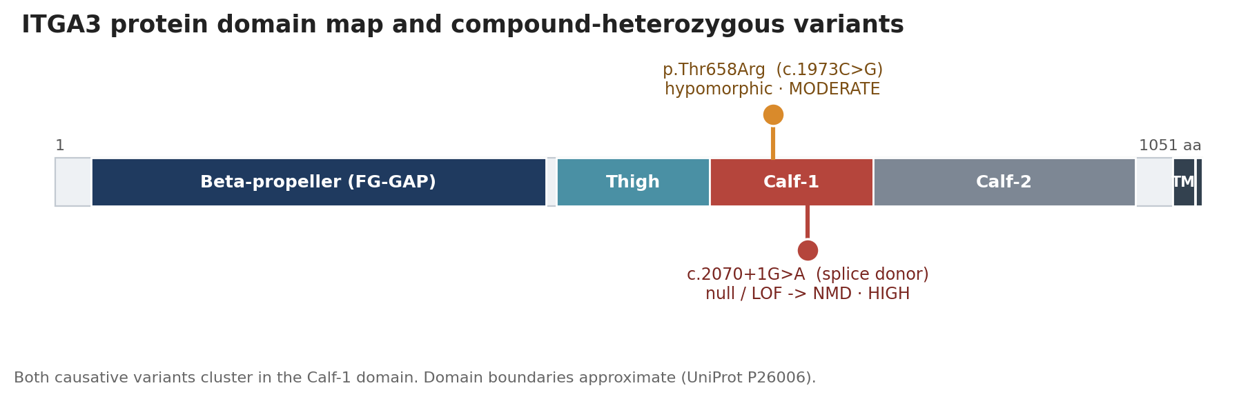
Figure 1. ITGA3 protein (NM_002204.4; UniProt P26006) with the two causative Calf-1 variants: the hypomorphic missense p.Thr658Arg and the null splice-donor c.2070+1G>A. Domain boundaries are approximate.
Supporting in-silico evidence for allele B included SIFT deleterious (0), PolyPhen-2 probably damaging (0.994), CADD 25.6, and REVEL 0.392. Zygosity and phase (in trans) derive from an independent variant-interpretation report and clinical genetics rather than from pipeline genotype calling; the compound heterozygosity is independently classified as Pathogenic / Likely Pathogenic. The predicted consequence is a marked reduction of functional α3β1 heterodimer with some residual protein from the hypomorphic allele, which is therapeutically relevant because it keeps protein-stabilization strategies on the table. A mechanistic literature anchor connects nephronectin (NPNT) signaling through ITGA3 to YAP1 stability and alveolar epithelial injury in fibrosis models (PMID 40444575), consistent with the epithelial-primary framing.
A second, genotype-independent epigenetic axis
The patient is homozygous reference (G/G) at rs35705950, so the canonical MUC5B common IPF variant is not carried. Despite this, both the MUC5B and TERT promoters show cohort-outlier hypermethylation on a conservative gate (islands with cohort_comparison=ALL and cohort_n at least 50): MUC5B z = +3.21 (delta-methylation +0.315) and TERT z = +2.77 (delta-methylation +0.243). This nominates a second, independent pathway toward the same clinical endpoints, mucin dysregulation and epithelial senescence, that would be invisible to standard variant-based IPF testing. The z-score cohort is small, so these are relative outlier metrics rather than calibrated p-values.
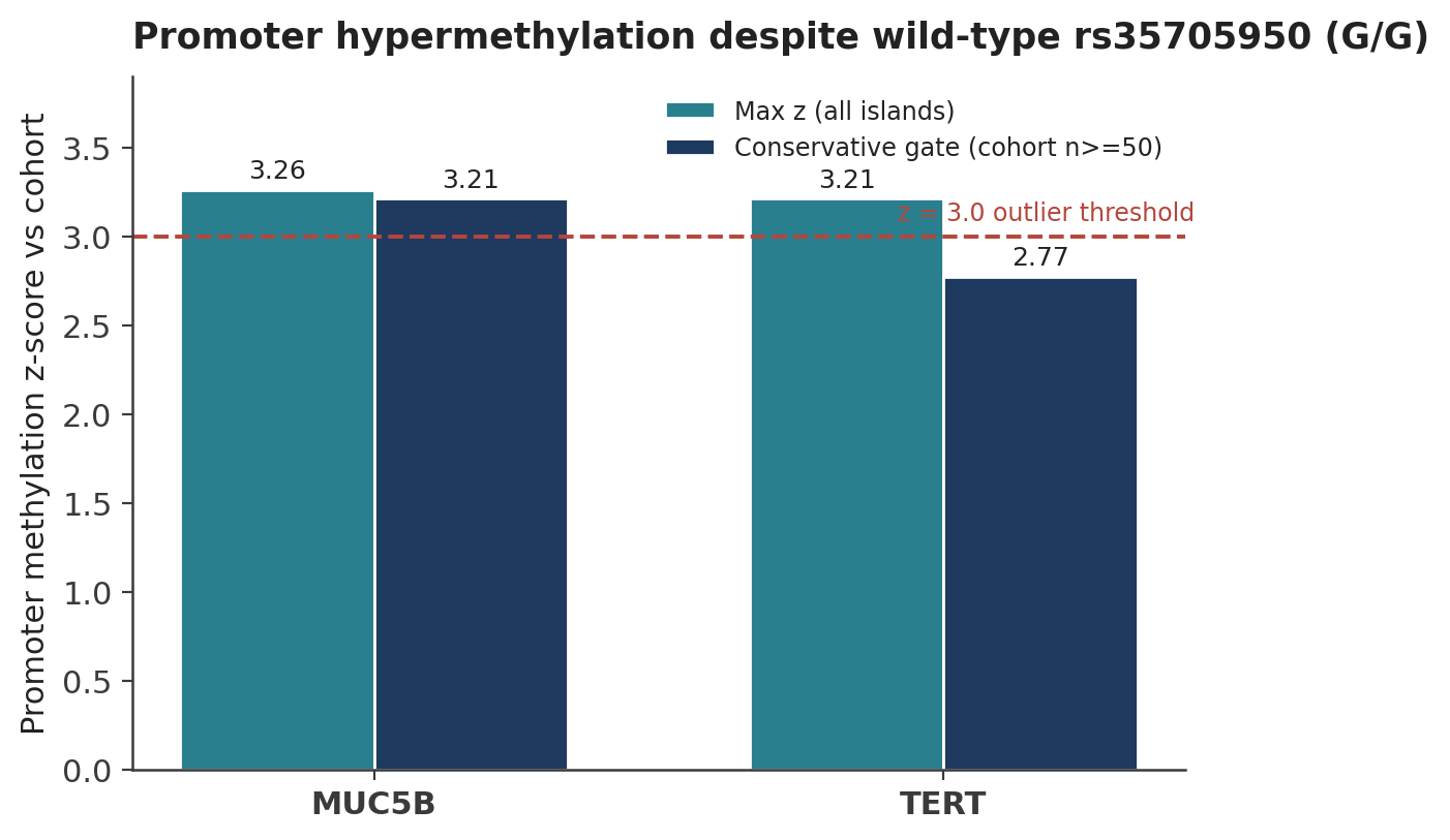
Figure 2. MUC5B and TERT promoter methylation z-scores versus the reference cohort, shown for all islands and for the conservative gate (cohort n≥50), against the z=3.0 outlier threshold. The patient is homozygous reference at rs35705950.
Plasma proteomics: epithelial-skewed and regulatory, not inflammatory
After LOESS batch correction, the plasma signature is dominated by epithelial-injury markers and is regulatory rather than pro-inflammatory. Headline signals (log2 fold change; assay-exchangeability permutation p, not genome-wide FDR): KRT19 +2.03, SPP1 (osteopontin) +1.55, and IL6 +1.96 elevated, with IL13 -1.97 and CCL18 -1.58 depleted. TGF-beta1 was not elevated in plasma (log2FC -0.23), consistent with tissue-compartmentalized rather than systemic fibrotic signaling. Because this is an n=1 comparison against a small reference pool, no individual protein reaches p < 0.05; signal class and direction are more trustworthy than any single call, and ITGA3 itself is not on the Olink panel, so protein loss is inferred rather than measured.
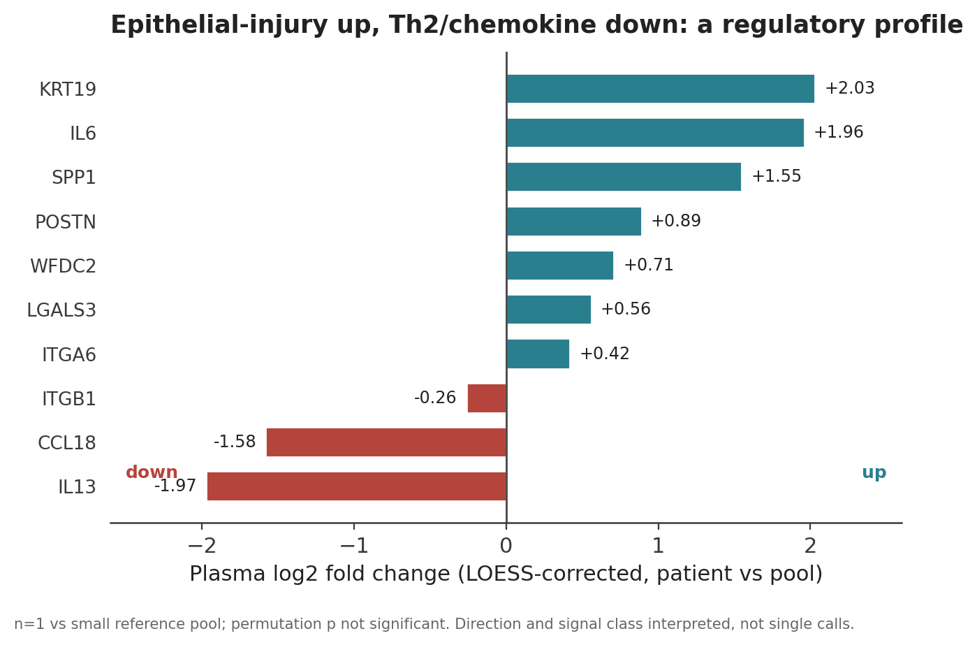
Figure 3. LOESS-corrected plasma proteome headline markers (log2 fold change, patient versus reference pool). Epithelial-injury markers rise while Th2 and chemokine markers fall. Values are n=1 and do not reach permutation significance; direction and signal class are interpreted.
Peripheral immune profile (scRNA-seq PBMC)
Two PBMC samples drawn at the same 2025 visit showed a quiescent CD8 T-cell expansion (approximately 3.7-fold versus reference, but with low granzymes and low exhaustion markers), an active Treg/Th17 axis concordant with the regulatory plasma phenotype, and a novel approximately 2.2-fold enrichment of ITGA6-positive NK cells not predicted by literature. ITGA3 expression in PBMCs was low across all cell types, as expected, since PBMCs are not the relevant compartment.
Hair-follicle long-read transcriptomics
Two independent long-read platforms confirmed ITGA3 isoform disruption in a low-ITGA3 surrogate tissue. Iso-Seq detected 26 patient-private ITGA3 isoforms with zero isoforms shared with control, consistent with LOF-induced isoform disruption rather than complete silencing. Complete Genomics RNA (after library-size normalization) showed upregulation of fibrosis markers COL1A1 and POSTN and loss of ESRP1. Epithelial-to-mesenchymal transition (EMT) markers were discordant between the two hair-follicle platforms and, importantly, EMT was not established in hair follicle, indicating that EMT in this patient is lung-tissue specific.
Longitudinal spatial lung analysis (2017 versus 2022)
The paired spatial biopsies are the same patient’s tissue five years apart rather than matched regions from one specimen, so sampling location and biological state are confounded; directions and deltas are interpreted rather than raw percentages as calibrated counts. Within that frame, the dominant longitudinal signal is a major immune reorganization.
| Cell type (PhenoCycler protein) | 2017 % | 2022 % | Delta (pp) |
|---|---|---|---|
| T_cell | 3.4 | 26.7 | +23.3 |
| Myofibroblast | 17.8 | 4.5 | -13.2 |
| Treg (cluster gate) | 8.9 | 0.0 | -8.9 |
| Neutrophil | 2.2 | 9.2 | +7.0 |
| Basal_epithelial | 1.6 | 0.1 | -1.5 |
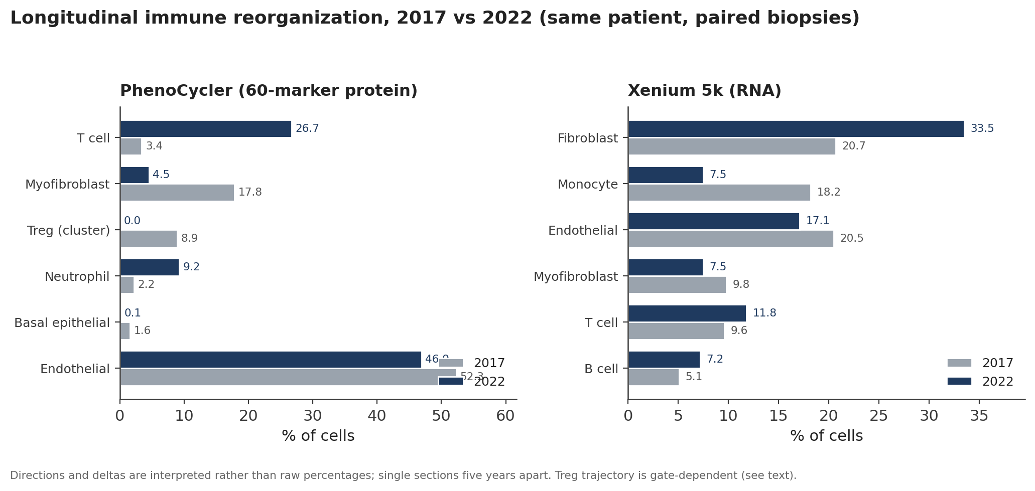
Figure 4. Longitudinal spatial cell-type composition at the 2017 and 2022 biopsies, by PhenoCycler protein (left) and Xenium RNA (right). The dominant signal is tissue T-cell expansion alongside airway basal-epithelial loss and stable-to-decreasing myofibroblast burden.
Key readings: an approximately eight-fold tissue T-cell expansion (Xenium shows the same direction, 9.6% to 11.8%); airway basal-epithelial collapse (1.64% to 0.12%), read as loss of the airway basal-cell niche that depends on α3β1; a neutrophilic component consistent with BOS; and a myofibroblast decrease across both spatial modalities (Xenium 9.8% to 7.5%), which under a constrictive-bronchiolitis lens is expected rather than evidence of inactive disease. Xenium differential expression confirmed ITGA3 downregulation in fibrotic zones (log2FC -1.13, adjusted p = 2.0 x 10^-5) alongside canonical fibrosis genes (FN1, COL1A1, COL3A1, ACTA2, POSTN) up and epithelial markers (SFTPC, AQP3) down, with intermediate-EMT cells enriched in fibrotic zones (Fisher OR 2.10) and confirmed at protein level by PhenoCycler (OR 2.36).
A specific longitudinal uncertainty (Treg trajectory). PhenoCycler cluster-based gating shows Treg collapse (8.9% to 0.04%), but rule-based FOXP3/CD4 gates on the same slides invert the direction (approximately 0.13% to approximately 8%). Blood TSDR supports current peripheral Treg deficiency but cannot adjudicate the lung-tissue 2017-to-2022 trajectory. This gating discrepancy is the principal unresolved construct-validity tension in the analysis.
Histomorphometry. Automated collagen-like area fraction (a blue-dominance heuristic, not an Ashcroft score) actually decreased between timepoints on cassette-matched Masson sections (2017 A2 11.63% to 2022 A3 10.64%), with a concordant EVG trend, so there is no histologic progression by these particular metrics.
Integrated causal model and hypothesis tests
All modality outputs were evaluated against a 247-edge, 106-node prior DAG. Of 231 patient DAG edges, 43 were directly confirmed and none were contradicted. The prior graph is anchored in alveolar UIP biology, so the confirmed subgraph is richer along the epithelial and immune axes than along the alveolar-myofibroblast axis, an asymmetry that is expected under the BOS reframe.
| Hypothesis | Verdict | Confidence |
|---|---|---|
| H1: Epithelial-primary fibrosis endotype | Partially supported | Low |
| H2: Decoupled IL-20 epithelial repair (receptors present, ligands low) | Supported | High |
| H3: Epigenetic axis (MUC5B/TERT hypermethylation) | Supported | Moderate |
| H4: EMT state, cross-modality | Partially supported | Low |
| H5: Immune remodeling (ITGA6 context plus CD8/Treg) | Partially supported | Moderate |
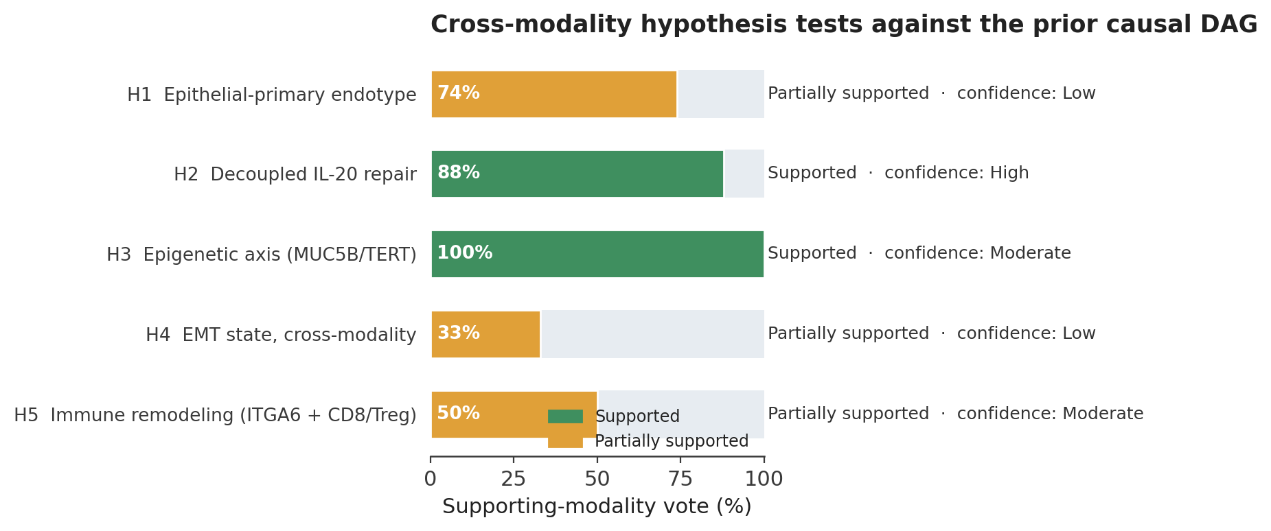
Figure 5. Cross-modality hypothesis tests (H1 to H5) scored as a supporting-modality vote against the prior causal DAG, colored by verdict with confidence annotated.
Cross-platform fold-change concordance was strongest for ACTA2 and ITGB1 (concordant up across all three RNA platforms); fibrosis genes (FN1, COL1A1, POSTN) were discordant between hair follicle and lung, reflecting tissue-of-origin biology rather than platform failure.
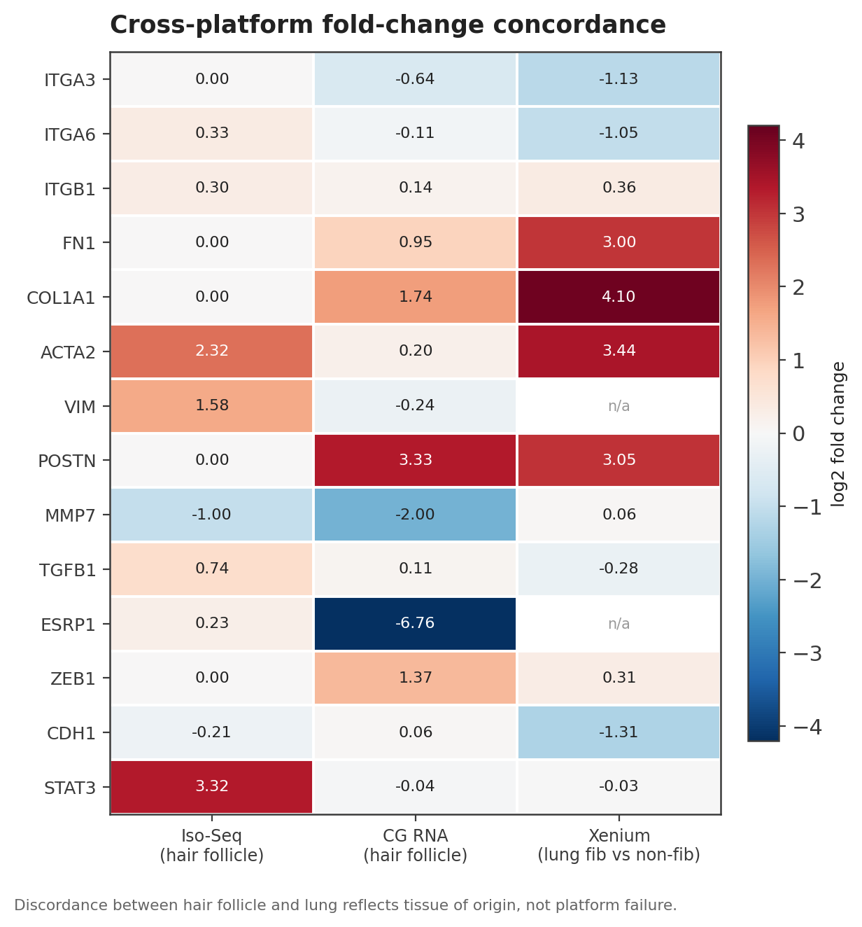
Figure 6. Gene-level log2 fold-change concordance across Iso-Seq and Complete Genomics RNA (hair follicle) and Xenium (lung, fibrotic versus non-fibrotic). Cells marked n/a were not measured on that platform.
Potential treatment insights
None of the following is a clinical recommendation; all require physician review, and every candidate is hypothesis-level given the n=1 design. Three independent lines of evidence were combined: patient pharmacogenomics, a curated map of PF-relevant drugs onto the confirmed DAG, and an unsupervised signature-reversal query.
- Phenotype-matched (clinician-priority). Given the small-airway / BOS-like phenotype, cGVHD-BOS-relevant agents are prioritized: belumosudil (FDA-approved ROCK2 inhibitor for chronic GVHD including BOS), fasudil (pan-ROCK), and axatilizumab (anti-CSF1R). The ROCK axis is cross-validated because fasudil also emerged independently from signature reversal. The repurposing rationale here is phenotype matching, not shared etiology, since the patient had a kidney (not allo-HSCT) transplant.
- IL-6 blockade. Tocilizumab and siltuximab match the elevated plasma IL-6 signal (log2FC +1.96).
- DNMT inhibitors. Azacitidine and decitabine directly target the MUC5B/TERT hypermethylation axis, which no approved PF drug addresses; decitabine also appears independently in signature reversal, making this the strongest cross-validated repurposing signal.
- Mechanism-of-action tests for H2. Recombinant IL-22 (F-652) or IL-20-family agonism directly test the decoupled-repair hypothesis (receptors expressed in epithelium, ligands low).
- De-prioritized. Blunt pan-TGF-beta and alpha-v-integrin ligand blockade is down-weighted given recent program safety setbacks (for example the discontinuation of bexotegrast in BEACON-IPF, NCT04396756; and the historical BG00011/STX-100 anti-alpha-v-beta-6 program), favoring upstream contextual modulation (ROCK, PDE4) instead.
Pharmacogenomic constraints (PharmCAT). Actionable calls include HLA-B*57:01 positive (abacavir contraindicated), SLCO1B1 decreased function (avoid high-dose simvastatin), CYP2C19 intermediate metabolizer (clopidogrel reduced efficacy; increased PPI exposure), CYP2C9 with VKORC1 (warfarin dose reduction if anticoagulation is needed), and CYP3A5 *3/*3 poor metabolizer (standard lower tacrolimus dosing appropriate). Any new candidate must be evaluated against transplant-mandated tacrolimus and mycophenolate, which cannot be freely modified for lung disease.
Discussion
What makes this case and approach unique. This is, to our knowledge, one of very few longitudinal, seven-modality molecular portraits of ITGA3-LOF pulmonary disease, and it is analyzed with an explicit causal-DAG framework rather than a modality-by-modality narrative. Two design choices carry most of the novelty. First, mapping every finding onto a prior causal graph converts a scattered n=1 dataset into a traceable, patient-confirmed subgraph in which each mechanistic claim is tied to specific modality evidence and each unresolved edge is explicitly logged. Second, the analysis reframes the disease from alveolar UIP to a small-airway, constrictive-bronchiolitis (BOS-like) endotype, a reframe that independently reconciles otherwise puzzling observations (airway basal-cell loss as a primary lesion, low and decreasing myofibroblast burden, and a dominant T-cell surge) into a single internally consistent picture that matches the histopathology.
How it expands current knowledge. The case adds two mechanistic points beyond the expected α3β1 adhesion defect. It documents a second, genotype-independent epigenetic axis (MUC5B and TERT promoter hypermethylation despite a wild-type risk allele), and it surfaces unexpected immune biology, including a quiescent CD8 expansion and an ITGA6-positive NK enrichment, that would not have been predicted from the germline lesion alone. In a single-patient study, unexpected findings that survive multi-modal cross-checking are arguably more informative than the expected ones, which are more vulnerable to confirmation bias.
Clinical implications. - Diagnosis: standard IPF genetic and inflammatory panels under-describe this disease; germline ITGA3 status, the epigenetic axis, and the BOS-versus-UIP distinction each change the picture. KRT19 and SPP1 emerged as the strongest plasma biomarkers and may be worth tracking longitudinally. - Prognosis: the plasma profile is regulatory rather than inflammatory, so routine inflammatory markers may underestimate disease activity, and the progressive small-airway obstruction is consistent with the severe, transplant-evaluation-level trajectory. - Treatment: reasoning shifts away from pan-TGF-beta ligand starvation and toward phenotype-matched BOS agents, IL-6 blockade, and DNMT-directed candidates, all bounded by transplant-mandated immunosuppression and the pharmacogenomic profile.
Limitations. This is a single patient, so all directional claims are patient-level and none is generalizable to IPF without replication. There is no direct functional validation of ITGA3 protein loss, which is inferred from genotype and downstream signals because neither the Olink nor the PhenoCycler panel includes ITGA3; a targeted western blot or mass spectrometry would close this loop. The two spatial timepoints are single sections five years apart with differing panels and fixation, so cell-type shifts describe available tissue rather than a guaranteed whole-lung trajectory, and the Treg longitudinal direction remains gate-dependent and unresolved. Reference pools for proteomics and methylation are small, no individual proteomic call reaches statistical significance, and the LINCS perturbations are dominated by cancer cell lines, so repurposing hits are hypothesis-generating only. Variant effect prediction would be tightened by LOFTEE and SpliceAI annotation, and state-versus-disease effects would be separable only with longitudinal blood.
Conclusion
- Compound-heterozygous ITGA3 LOF (one null splice allele plus one hypomorphic missense allele) is a confirmable, epithelial-primary driver of pulmonary fibrosis that standard IPF genetic testing does not capture.
- The disease is best framed as a small-airway, constrictive-bronchiolitis (BOS-like) endotype, not alveolar UIP; this reframe reconciles the spatial, histologic, and clinical evidence and reorients therapy toward phenotype-matched, cGVHD-BOS-relevant agents.
- A second, genotype-independent epigenetic axis (MUC5B and TERT promoter hypermethylation without the rs35705950 risk allele) nominates DNMT-directed intervention as a distinct, cross-validated therapeutic hypothesis.
- Beyond this patient, the case shows that mapping multi-modal data onto a prior causal graph can extract traceable, clinically actionable mechanism from a single rare-disease patient, provided every claim remains scoped to the n=1 evidence that supports it.
Be the first to comment on this publication.