Purpose
Background
Biallelic loss of ITGA3, encoding integrin α3, causes interstitial lung disease, nephrotic syndrome and epidermolysis bullosa (ILNEB), classically presenting as an infantile alveolar lung disease. However, how a single missense allele in this gene reshapes the α3β1 receptor is rarely characterised at the tertiary protein-structure level. Because α3β1 function depends on receptor conformation as well as expression, we use computational methods to ask whether T658R destabilises the receptor fold or instead changes its local shape and dynamics.
Case
We studied an adult man whose biallelic ITGA3 disease presented atypically as airway-predominant pulmonary fibrosis resembling bronchiolitis-obliterans syndrome, in the setting of kidney transplantation and on chronic immunosuppression. His genotype was compound heterozygous in trans: a paternal splice-donor allele predicted to undergo nonsense-mediated decay, and the maternal missense allele T658R, in which threonine at position 658 is replaced by arginine. The cellular α3β1 pool is therefore expected to depend largely on a single altered α3 allele. T658R lies in the region of the α3 leg that binds CD151, a tetraspanin partner that stabilises the adhesion complex.
Molecular findings.
Because this was a single, chronically immunosuppressed patient, changes in his blood-protein and gene-expression data could not be separated from medication effects. We therefore focused on the protein itself and asked what T658R mutation does to α3β1 on the tertiary protein structure level. We used three complementary computational approaches: FoldX, to estimate whether the mutation destabilises the folded protein; AlphaMissense a machine-learning method, to score the likely pathogenicity of the missense change; and molecular dynamics (MD), using repeated all-atom simulations of an AlphaFold model of the α3β1 ectodomain, as no experimental structure is available. Wild-type and mutant receptors were simulated separately. Neither FoldX nor AlphaMissense showed a significant difference between wild type and the mutant. The simulations, by contrast, showed a clear conformational effect: T658R bent the α-genu hinge, the α-knee between the receptor headpiece and lower legs, from about 139° in the wild type to about 130° in the mutant, a roughly 9° sharper bend, and it loosened the α3 leg. The folded β-sheet domains on either side of this hinge retained their shape, with no loss of secondary structure. Rather than producing one dominant new interaction, the introduced arginine reshaped local contacts around residue 658. Together, the FoldX, AlphaMissense and MD results suggest that T658R leaves the fold intact while introducing conformational changes: sharper α-genu bending and loosening of the α3 leg.
Clinical implications.
The main value of this analysis is that it provides a computational hypothesis for how the T658R mutation could affect the observed ILNEB phenotype, in particular with regards to the tertiary protein structure of α3β1. The next experiments should test whether the T658R-induced changes affect receptor surface expression, glycosylation, CD151 binding or activation state. If this model is confirmed, future therapeutic work should focus on the mechanisms that control α3β1 conformation, trafficking and interaction with CD151, rather than on generic fold stabilization alone.
Keywords: ITGA3; ILNEB; integrin α3β1; T658R; α-genu; molecular dynamics; CD151; pulmonary fibrosis; variant effect
Introduction:
Cells anchor to the scaffold around them through adhesion receptors called integrins; when one such receptor is altered, the cell can adhere and signal abnormally. Integrins link the extracellular matrix to the cytoskeleton and are allosteric, signalling by changing shape rather than only by binding. The laminin-binding integrin α3β1, whose α subunit is encoded by ITGA3, is a principal adhesion receptor of epithelial basement membranes. Together with its laminin-332 substrate and the tetraspanin CD151 (a four-pass membrane protein that laterally associates with α3β11 and stabilises the adhesion complex), it organises the epithelial-matrix interface. Thus, α3β1 function depends not only on expression but also on receptor conformation.
Biallelic loss of ITGA3 causes a syndrome of interstitial lung disease, nephrotic syndrome and epidermolysis bullosa (ILNEB), in which the three features co-occur, classically in infants.2 The phenotypic spectrum has since broadened to include milder, later-presenting compound heterozygous cases,3–4 and, more rarely, survival to adulthood.5 Across these reports, the laminin-332/α3β1/CD151 module is the shared point of failure. These cases are rare, and the link between a particular α3 substitution and its cellular consequence is seldom resolved at the tertiary protein structure level.
The patient we describe is an adult man with severe, progressive pulmonary fibrosis caused by biallelic ITGA3 mutations. His course was atypical. After kidney transplantation and while on chronic immunosuppression, his lung disease was airway-centred, resembling bronchiolitis-obliterans syndrome with lymphocytic bronchiolitis and relatively sparing of the alveolar compartment. This pattern differs from the alveolar, infantile presentation classically described for ITGA3-related disease. His genotype is compound heterozygous in trans: a paternal splice-donor allele predicted to undergo nonsense-mediated decay (NMD), and the maternal missense allele T658R, which sits in the calf-1 domain of the α3 subunit (Fig. 1, shown in detail in Fig. 3).
This setting makes the conformational mechanics of the receptor directly relevant. The integrin ectodomain switches between bent, low-affinity and extended, high-affinity states through a large, slow transition organised around the α-knee, also referred to as the α-genu (Fig. 1), which lies between the headpiece and the lower-leg (calf) domains. Force transmitted through the cytoskeleton across this hinge can switch the receptor’s affinity sharply.6–9 The α-genu is a deformable hinge, as shown by structural and simulation studies,10–11 and residue 658 lies in the calf-1 domain (Fig. 1). A substitution that biases this hinge could therefore alter receptor behaviour while leaving the fold intact and the protein expressed.
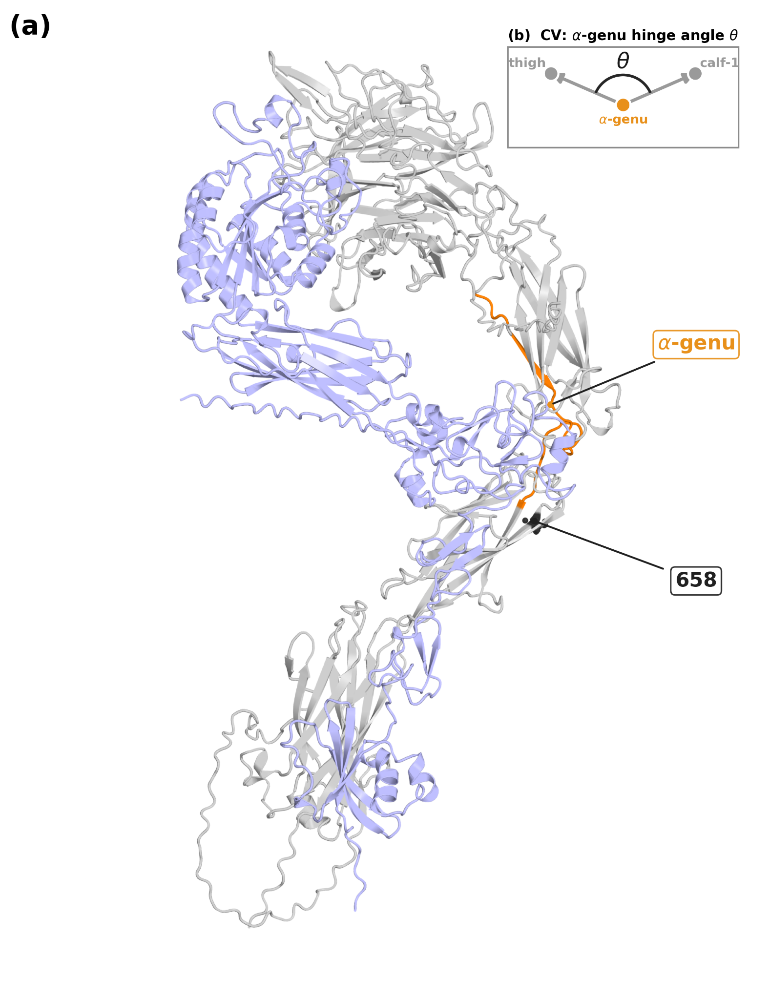
Figure 1. he α3β1 ectodomain and the α-genu hinge angle. (a) The simulated apo wild-type α3β1 ectodomain dimer (an AlphaFold-derived model). The α3 subunit is light grey, the β1 subunit pale blue, and the α-genu hinge region (α3 residues 600–625) is highlighted in orange; the disease residue 658, in the calf-1 domain just below the α-genu, is marked. (b) The α-genu hinge angle θ, defined as the angle at the α-genu-domain Cα centre of mass (COM) between the vectors to the thigh and calf-1 COMs; this is the main quantity we track in the simulations.
In ultra-rare disease, variant classification often leaves the structural mechanism unresolved. Such classification rarely shows how the change actually alters the protein’s shape and movement. With only one patient (n=1), there is no comparison group, so population-genetic evidence of risk is unavailable. The patient has also received several immunosuppressive drugs since his kidney transplant, so any change in his blood proteins or gene expression could reflect medication rather than the variant, and the two cannot be separated in a single treated person.
At the tertiary protein structure level, an open question remains: does α3β1 drive lung fibrosis or does it protect against it? Epithelial α3β1 has been reported as pro-fibrotic, linking β-catenin and Smad signalling to myofibroblast formation,12 yet α3β1 with CD151 has also been reported to protect the epithelium,13 and a nephronectin-ITGA3-YAP1 axis has recently been described in idiopathic pulmonary fibrosis;14 Here, we focus on a narrower question: how the T658R substitution affects α3β1 structure and dynamics.
Here we characterise T658R at the level of the α3β1 protein, following the workflow in Fig. 2. We combine folding free-energy estimation (FoldX), machine-learning pathogenicity prediction (AlphaMissense), and multi-replica all-atom molecular dynamics simulation (MD) of the α3β1 ectodomain to compare the wild-type and T658R receptors. We also compare T658R with other experimentally characterised calf-1 mutants reported in the literature15 (Fig. 3). The variant T658R does not destabilise the fold, and the AlphaMissense classifier cannot decide either way, yet it changes the receptor’s motion: it biases the α-genu toward a more acutely bent state and loosens the α3 leg, reshaping local contacts around the mutated residue while the calf-1 domain keeps its folded shape. T658R also disrupts the local N-linked glycosylation sequon Asn656-Val657-Thr658. GlyGen reports Asn656 as an experimentally observed glycosylation site with published glycoproteomic mass-spectrometry evidence16, so T658R is predicted to remove Asn656 glycosylation.
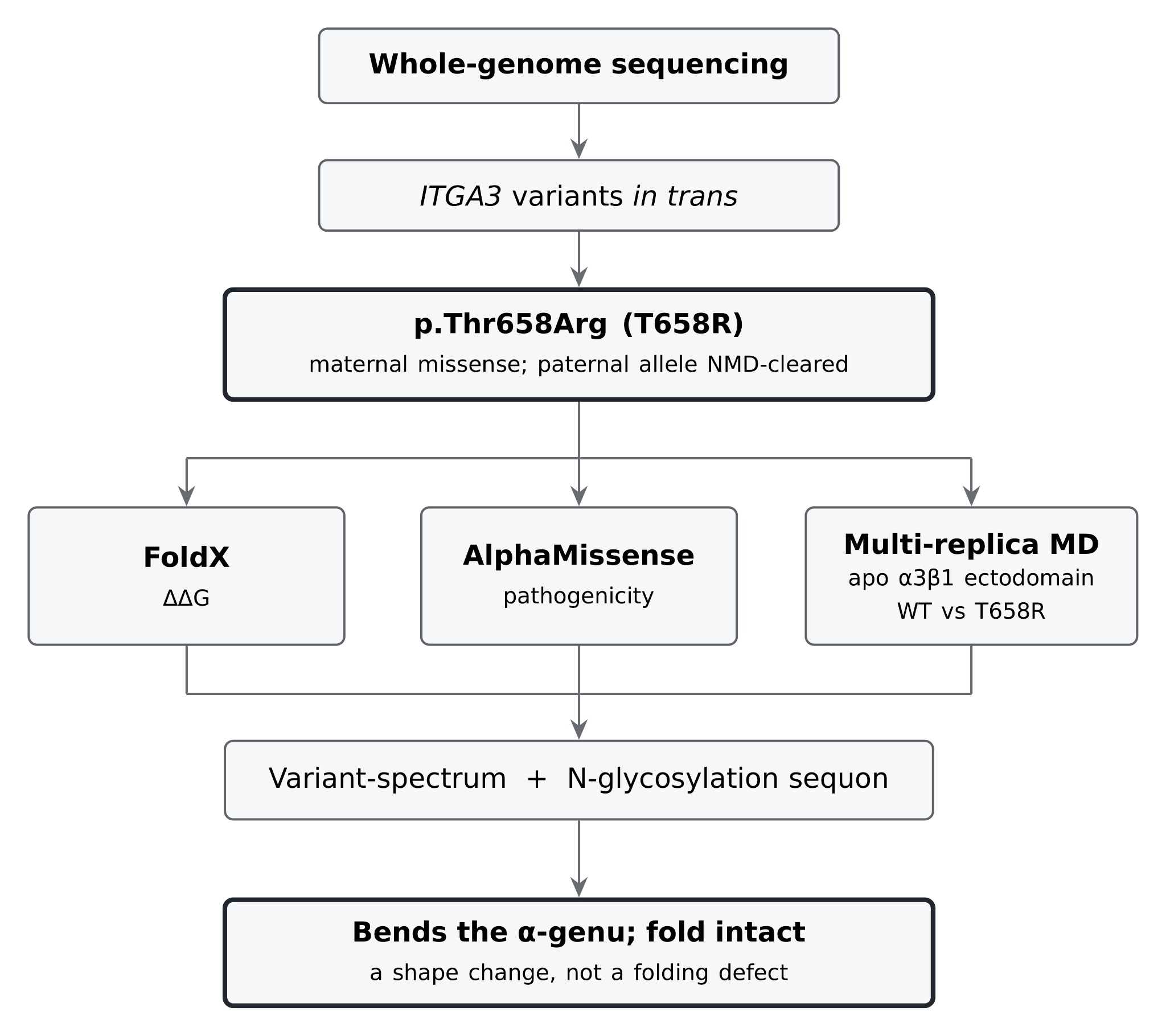
Figure 2. Analytic workflow. The maternal T658R allele is characterised at the protein level with FoldX, AlphaMissense and multi-replica molecular dynamics of the apo α3β1 ectodomain (wild type vs. T658R), then T658R is compared with known calf-1 variants and assessed for loss of the Asn656-Val657-Thr658 N-glycosylation sequon.
Case presentation:
Patient and family history.
The patient is an adult man, in his early thirties at the time of study, with biallelic ITGA3 disease. In early adulthood he developed end-stage renal disease and received a kidney transplant from a related living donor, with no subsequent allograft rejection. He has remained on maintenance immunosuppression since transplantation. The renal findings are consistent with the nephrotic component of the interstitial lung disease, nephrotic syndrome and epidermolysis bullosa (ILNEB) phenotype associated with ITGA3.2 he family history fits the compound heterozygous genotype in trans: the parent carrying the splice-donor allele has a mild ectodermal phenotype (enamel and skin), whereas the parent carrying the missense allele is unaffected, consistent with each single allele being tolerated on its own and disease arising only when both are inherited.
Pulmonary presentation.
The defining problem is severe, progressive pulmonary fibrosis. The disease was airway-predominant and presented in adulthood, with progressive exertional dyspnoea and supplemental-oxygen dependence. Pulmonary-function testing showed a severe mixed obstructive–restrictive ventilatory defect, with markedly reduced forced expiratory volume in 1 s, forced vital capacity and diffusing capacity (FEV1 25%, FVC 37% and DLCO 42%). The clinical and physiological picture resembled bronchiolitis obliterans syndrome (BOS) rather than a usual interstitial pneumonia pattern.
Diagnostic workup.
The diagnosis combined imaging, lung histology and genomic testing. Cross-sectional imaging and physiology indicated small-airway disease with relative sparing of the alveolar compartment. Two surgical lung specimens were obtained at different times as wedge resections during management of spontaneous pneumothorax (with bleb resection and pleurodesis). These sampled peripheral lung at the pneumothorax site and were therefore not primarily diagnostic of the airway-centred disease process. Whole-genome sequencing established the molecular diagnosis (detailed in the Results), showing two ITGA3 variants in trans.
Pathology.
Histology showed an airway-centred fibrotic process: bronchiolitis obliterans with lymphocytic bronchiolitis, and comparatively preserved alveolar architecture, a pattern resembling chronic graft-versus-host-like airway disease, even though the patient received a solid-organ transplant (kidney), not a haematopoietic stem-cell, transplant. A minor salivary gland biopsy showed extensive fibrofatty replacement with sicca symptoms, indicating that the fibrosing process is not confined to the lung. Together, the findings describe systemic, airway- and exocrine-predominant fibrosis on a background of ITGA3 deficiency and chronic immunosuppression.
Available data extracted from the patient.
Seven datasets had been generated for this patient: long-read whole-genome sequencing (blood), DNA methylation (blood), plasma proteomics (Olink), single-cell RNA-seq (blood), spatial transcriptomics (Xenium, lung), spatial protein imaging (PhenoCycler, lung), and long-read RNA-seq (Iso-Seq, hair follicle). Of these, only the whole-genome sequencing was used here, for variant calling and phasing. The other datasets were not analysed as results. In a single, chronically immunosuppressed transplant recipient, omics cannot be separated from drug and transplant effects, so the study is centred on what the maternal missense allele does to the protein.
Results: Molecular Analysis and Interpretation
T658R shows no clear signal of fold damage or pathogenicity.
Whole-genome sequencing identified two ITGA3 variants in trans (Table 1): a paternal splice-donor allele predicted to be removed by nonsense-mediated decay, and the maternal missense allele T658R, from which most available α3 subunit is expected to derive. Before simulation, we used two fast tools to ask whether the change destabilises or clearly damages the protein. To test folding stability we used FoldX, an empirical calculator that estimates how a mutation changes a protein’s folding free energy. A protein’s folding free energy (ΔG) measures how strongly it prefers its correctly folded shape. FoldX reports the mutation-induced change (ΔΔG, the mutant value minus the wild-type value): a large positive ΔΔG means the fold is destabilised, and a value near zero means it is not. For T658R the change was small on both structures: ΔΔG = −0.37 kcal/mol on the isolated α3 subunit and −0.01 kcal/mol on the assembled α3β1 ectodomain (means of three runs each). FoldX treats any ΔΔG between about −0.5 and +0.5 kcal/mol as no change in stability. Both values therefore support a neutral effect, the slightly negative −0.37 value remains within the method’s uncertainty thresholds. Because residue 658 lies in the calf-1 leg, far from the α3–β1 interface, the mutation does not alter β1’s local environment. Consistent with this, the two ΔΔG estimates (for isolated α3 subunit and assembled α3β1 ectodomain) agree. Any functional defect therefore is more likely to reflect local conformational effects rather than overall folding thermodynamics.
As another independent sequence-based check on pathogenicity, we used AlphaMissense, a machine-learning tool that scores how likely each possible missense change in the human proteome is to cause disease, from 0 (benign) to 1 (pathogenic). Its authors fixed two cut-offs by calibration against clinically classified (ClinVar) variants: “likely benign” below 0.34 and “likely pathogenic” above 0.564, with scores in between labelled “Ambiguous”. T658R scored 0.4144, which is within the Ambiguous middle range. AlphaMissense therefore did not classify the variant as either likely benign or likely pathogenic. By contrast, the other charged or bulky substitutions at position 658 (T658D/E/H/K) scored near 0.6, in the pathogenic band.
| Allele | Position (GRCh38) | HGVS / protein | GT | Consequence | Effect |
|---|---|---|---|---|---|
| Paternal | chr17:50077122 | c.2070+1G>A | 1|0 | splice-donor | null; NMD-cleared |
| Maternal | chr17:50077024 | c.1973C>G, p.Thr658Arg | 0|1 | missense | expressed |
Table 1. The biallelic ITGA3 genotype (compound heterozygous, in trans; shared phase set PS=49433653, opposite blocks). NM_002204.4 is the MANE Select transcript. NMD, nonsense-mediated decay.
T658R sits in the CD151-binding region of calf-1.
α3β1 interacts with CD151, a small four-pass membrane protein (a tetraspanin) that binds the α3 leg and holds the adhesion complex together, so a change inside that binding region could affect this binding. Residue 658 lies in the calf-1 domain, within the α3 region that binds CD151 (residues 570–705, identified experimentally with α6/α3 chimeric integrins and chemical cross-linking; Fig. 3a).1 In the wild type, it is a neutral threonine, whose side chain points inside the α3 leg and away from the α3-β1 interface, so the mutation acts within α3. Residue 658 sits within the CD151-binding surface of α3. Therefore, replacing threonine with a larger, positively charged arginine (Fig. 3b,c) places a positive charge inside the CD151-binding surface and changes the local electrostatic profile, which could weaken CD151 recognition.
Previously published data on neighbouring Arg628 suggests that, in calf-1, the severe defect is driven by backbone disruption rather than charge change.15 On the other hand, the T658R mutation keeps the backbone intact. Substitutions at Arg628 reported by Yamada and Sekiguchi make this clear (Table 2): the disease-associated variant R628P introduces a proline that breaks the local β-sheet and cannot bind CD151, whereas the engineered charge-only substitutions R628Q and R628E behave like the wild type.15 T658R changes the charge but keeps the backbone, so we would not expect the same CD151 binding loss and endoplasmic-reticulum retention that make R628P pathogenic.
| Variant | Backbone | CD151 association | Trafficking |
|---|---|---|---|
| R628P15 (disease) | broken (proline) | abolished | ER-retained |
| R628Q / R628E15 (controls) | intact | normal | normal |
| T658R (this study) | intact | unknown | unknown |
Table 2. T658R compared with substitutions at calf-1 residue Arg628, all from Yamada and Sekiguchi.15 R628Q and R628E are engineered controls (charge changed, backbone kept), not disease variants. For T658R the backbone is intact (a buried side-chain charge, not a backbone break) and its effect on CD151 was not measured. ER, endoplasmic reticulum.
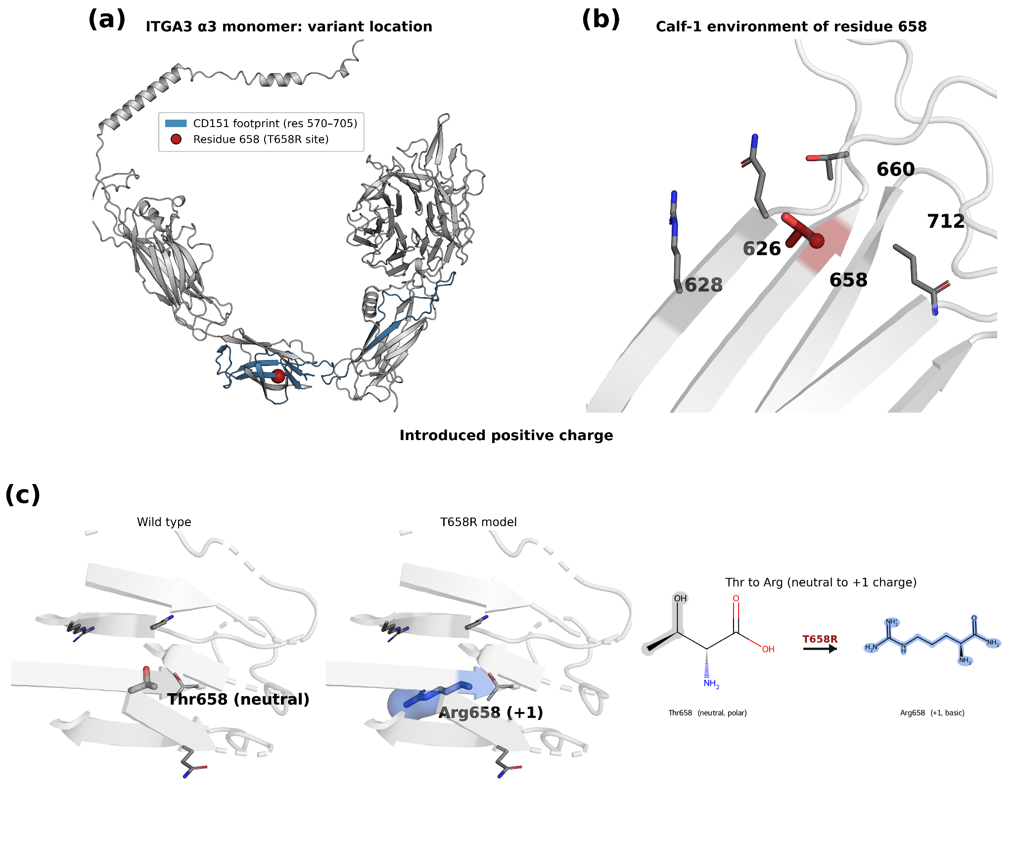
Figure 3. T658R in the CD151-binding region of calf-1. (a) The α3 monomer (AlphaFold P26006) with the CD151-binding region (residues 570–705) highlighted1 and residue 658 marked; positions 612 and 628 are shown for reference, of which only R628 is experimentally characterised.15 (b) The local environment of residue 658. (c) A qualitative view of the wild-type (Thr658) and mutant (Arg658, +1) site; the arginine adds one positive charge at a buried position.
With neither the FoldX stability calculation nor the AlphaMissense score giving a clear answer, and with published Arg628 data suggesting that charge change does not explain severe CD151-binding region defect, we turned to all-atom simulation to look directly for local changes in shape and motion to gain detailed insights into possible alteration of protein dynamics.
**The α-genu flexibility and bending changes in the "T658R" mutant.
**T658R closes the α-genu hinge, the clearest dynamic difference between the two receptors (Fig. 1 and Fig. 4). Over the 50 ns window, the α-genu angle fell from a per-replica mean of 139.1 ± 0.7° (wild type, n = 3) to 130.3 ± 2.8° (T658R, n = 8; mean ± standard deviation across replicas), a difference of about 8.7°. The mutant’s larger spread (±2.8° versus ±0.7°) is itself informative: the mutant not only bends more but samples a wider range of angles, matching the looser leg described below. To estimate the size of this difference, rather than only whether it is detectable, so we measured its effect size with Hedges’ g, a version of Cohen’s d corrected for small samples: g = −3.23, which is very large (values above 0.8 are conventionally called “large”). Consistent with this, the per-replica means, which are the statistically independent units, do not overlap at all: every T658R replica is more bent than every wild-type replica (a supporting exact permutation test on this non-overlap is reported in the Supplementary). Because the bent-to-extended transition occurs on microsecond-to-millisecond timescales9, far beyond 50 ns, each replica largely keeps the bending state it started in, which partly explains why the mutant angles scatter more; we therefore interpret this as a robust direction of effect
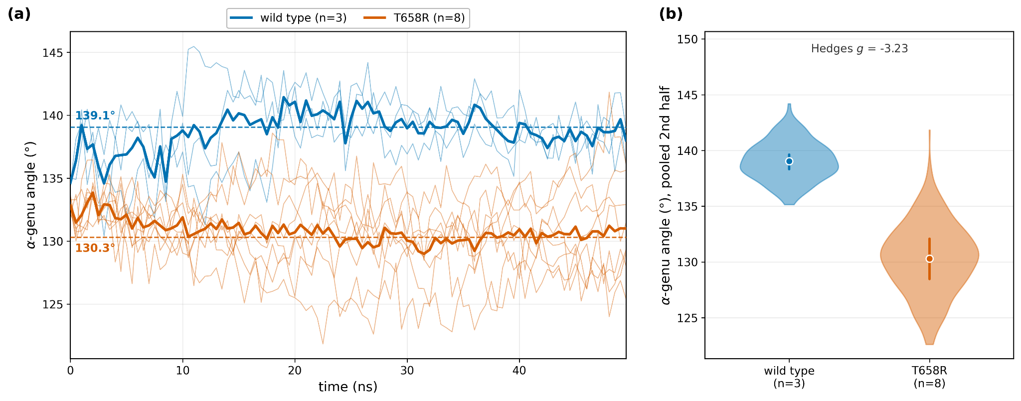
Figure 4. T658R bends the α-genu. (a) Per-replica α-genu angle versus time (thin lines) with per-condition means (bold) and the two arm means marked (139.1° wild type, 130.3° T658R). (b) Distribution of the pooled second-half angle per condition, showing the non-overlapping separation between wild type and T658R. Wild type (blue) n = 3, T658R (vermillion) n = 8.
Before interpreting the α-genu bend as a conformational signal, we checked that the receptor did not unfold. The root-mean-square deviation (RMSD) measures how far the atoms have moved, on average, from the starting structure; a rising RMSD means the overall shape or position is changing. The whole-ectodomain (α3+β1) Cα RMSD rose over most of the window and levelled off only near its end (wild-type final-frame mean 7.96 Å). The mutant settled at a higher value and fluctuated more (8.83 Å) (Fig. 5a). These large values reflect rigid-body motion, primarily the β1 leg swinging relative to α3, not unfolding, as is characteristic of integrin ectodomains.6–8 Within α3, the per-domain RMSD values are small (thigh, α-genu and calf-1 all about 1.2-1.9 Å), which confirms that the individual domains stay internally rigid and that the α-genu acts as a hinge between rigid bodies (Fig. 5b). The one domain where the mutant is modestly more mobile is the α-genu itself (1.91 versus 1.58 Å), consistent with an altered hinge.
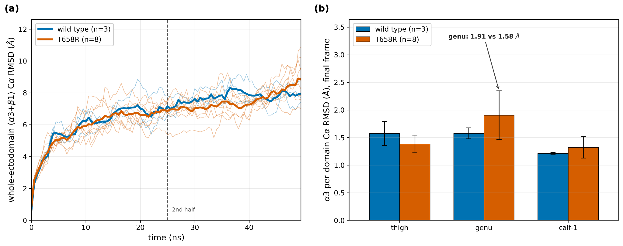
Figure 5. Receptor stability and domain rigidity. (a) Whole-ectodomain (α3+β1) Cα RMSD versus time, per replica (thin) and per-condition mean (bold); the dashed line marks the start of the second-half (25 ns) statistics window. The whole-ectodomain RMSD is dominated by β1 inter-chain motion. (b) Per-α3-domain final-frame RMSD (mean ± SD across replicas): thigh, α-genu and calf-1 are internally rigid in both arms, with the α-genu most elevated in T658R. Wild type n = 3, T658R n = 8.
The α3 leg loosens.
The mutation also increased flexibility across the α3 subunit. Where RMSD tracks the whole structure, the root-mean-square fluctuation (RMSF) measures how much each individual residue fluctuates around its own average position, so a higher RMSF means a more flexible residue. The mean Cα RMSF rose from 1.86 to 2.60 Å over the α3 subunit (the α3 chain within the dimer) and from 1.94 to 2.68 Å over the whole ectodomain (Fig. 6a). The extra flexibility was broad, not confined to residue 658. The T658R profile was higher than wild type along most of the α3 leg (Fig. 6a), and the per-residue difference (ΔRMSF, T658R minus wild type) was positive both at the mutated residue 658 (+0.75 Å) and across the surrounding calf-1 region (Fig. 6c); the whole ectodomain profile shows the same lower leg increase with the headpiece core unchanged (Fig. 6b, and Supplementary Fig. S2). The single largest per-residue difference is not at the mutation site but at a chain end (residue 23), the terminal fraying expected of an unconstrained chain terminus17 rather than an effect of the variant.
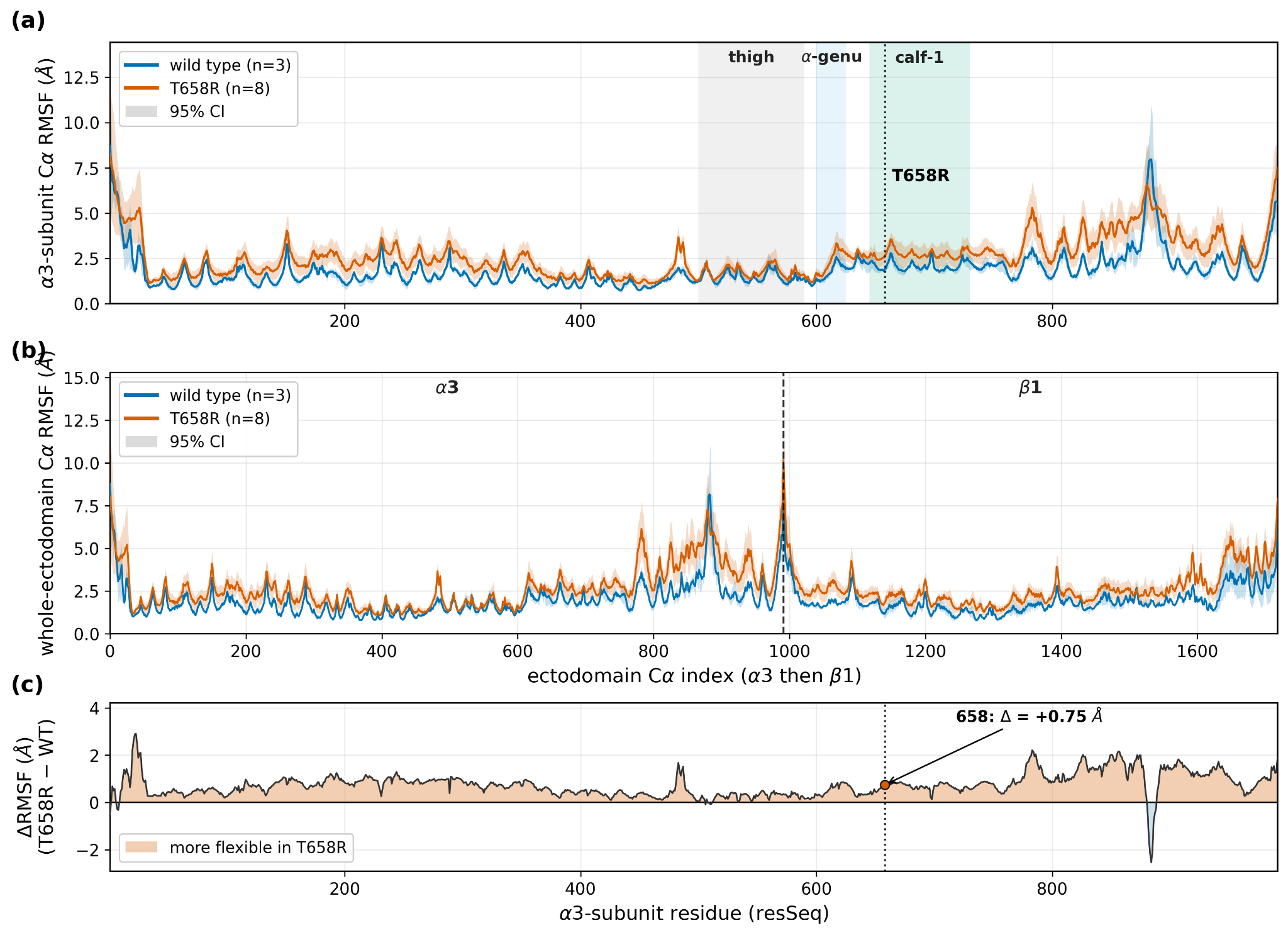
Figure 6. Per-residue flexibility (α3β1 ectodomain dimer). (a) α3-subunit per-residue Cα RMSF, wild type versus T658R, with 95% confidence interval ribbons across replicas; domain bands (thigh, α-genu, calf-1) shaded and the T658R site marked. (b) Whole-ectodomain per-residue Cα RMSF on a continuous Cα index, with the α3/β1 boundary marked. (c) Per-residue ΔRMSF along the α3 subunit (T658R minus wild type): positive across most of the leg, +0.75 Å at residue 658; the spike at the chain terminus is a fraying artefact. Wild type n = 3, T658R n = 8.
The mechanism at residue 658 causes reshaping of local contacts.
The larger arginine side chain points into the leg core rather than toward the solvent, so the substitution buries a positive charge inside the protein. There, the arginine side chain packed more tightly against its neighbouring residues and displaced the hydrogen bonds made by the wild-type threonine. This likely caused the calf-1 region to tighten the leg core and loosen the α-genu loop (Fig. 7), matching the broader rise in flexibility and the bend at the α-genu. We therefore interpret the contact changes as local reshaping around residue 658, not as evidence for one specific new stable interaction.
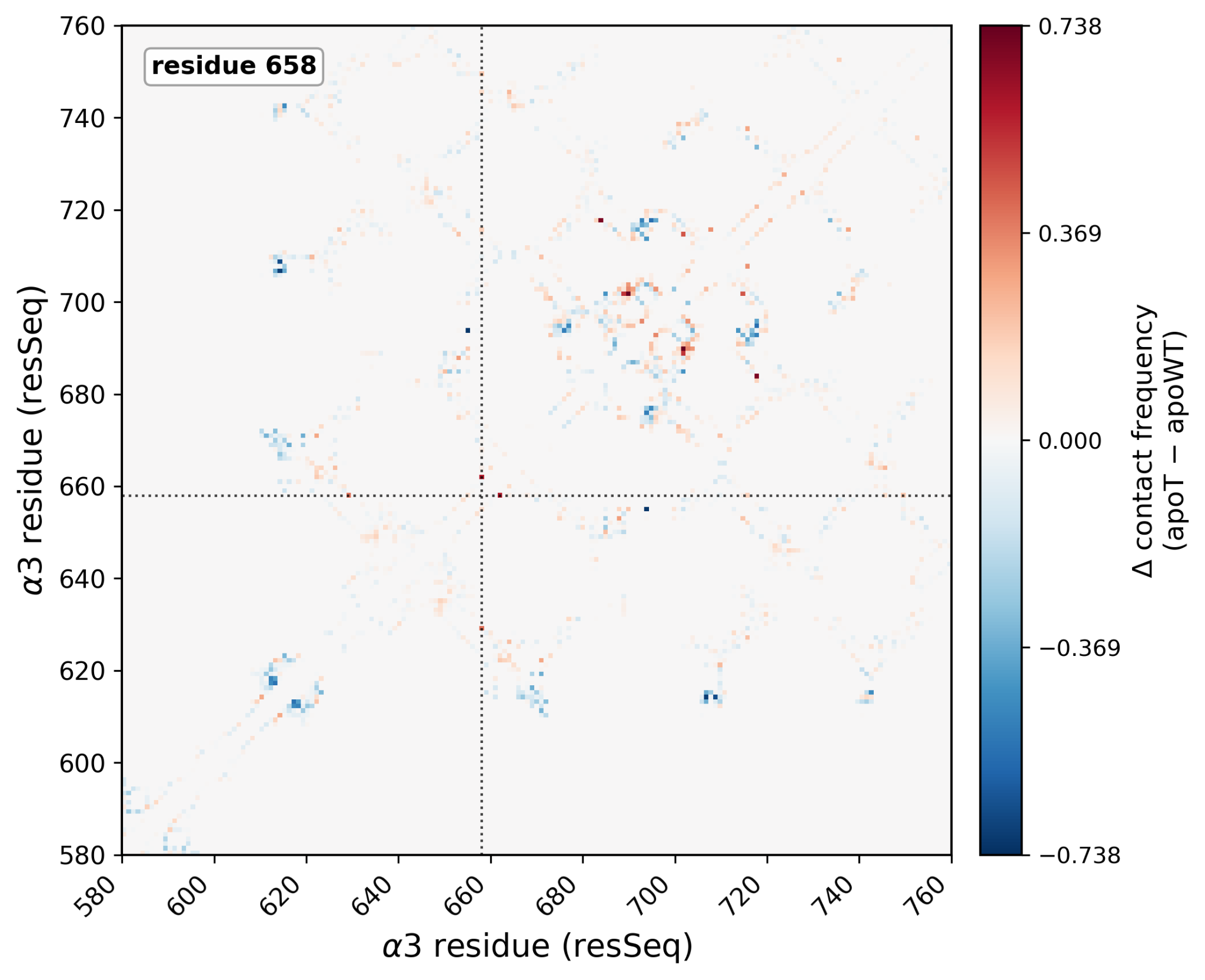
Figure 7. Local contact reshaping at residue 658. T658R minus wild-type contact-difference map over the α3 leg and α-genu region (α3 residues 580–760; red, contacts gained in T658R; blue, contacts lost); residue 658 is marked. The mutant gains compact calf-1-core contacts (for example residues 690–702 and 684–718) and loosens the α-genu loop (for example 655–694 and 614–707).
Methods, algorithms and tools:
**Patient variant and structural models.
**Phased whole-genome variant calls were provided for the patient, reported relative to the MANE Select transcript NM_002204.4 (GRCh38). The two ITGA3 variants share one phase set but lie on opposite haplotypes, which confirms a compound heterozygous genotype in trans. The paternal allele is a splice-donor change (c.2070+1G>A) predicted to trigger nonsense-mediated decay, and the maternal allele is the missense change c.1973C>G (p.Thr658Arg; T658R). Because most available α3 subunit is expected to derive from the missense allele, we built a T658R model from the wild-type α3β1 structure and studied the wild-type and T658R structures in silico. In the T658R model, Arg658 was treated as the standard protonated arginine side chain, with a cationic guanidinium group and fixed force-field partial charges. Buried arginine residues are generally expected to remain charged, but buried ionizable groups can be accommodated by local hydrogen bonding, water access and conformational reorganisation. These alternative responses were not directly sampled here.18–20
Because no experimental structure of the α3β1 ectodomain is available, all structural models were AlphaFold predictions. The α3 monomer was taken from the AlphaFold model of human integrin α3 (UniProt P26006),21 For the dynamics we used a model of the complete α3β1 ectodomain (α3 residues 1–991, β1 residues 1–728; the transmembrane and cytoplasmic segments were excluded so that the soluble ectodomain could be simulated in water) derived from an AlphaFold322 prediction, retaining seven structural metal ions at the canonical β-propeller domain, MIDAS (metal-ion-dependent adhesion site), ADMIDAS (the adjacent metal site) and SyMBS (synergistic metal-binding site), and at the α-genu site (Supplementary Information, Simulation parameters). The wild-type and T658R systems therefore differed only at residue 658, carrying threonine or arginine, respectively.
**Force field and MD protocol.
**All-atom molecular dynamics were run in OpenMM 8.423 with the AMBER ff14SB protein force field24 and the TIP3P water model.25 The structural metal ions were parameterised with the Li–Merz 12-6 ion–oxygen-distance set for divalent cations.26 Each system was solvated in a rhombic-dodecahedral box with 1.0 nm solute padding, neutralised, and brought to 150 mM NaCl, which gave about 1.02 million atoms. Electrostatics used particle-mesh Ewald (PME)27 with a 1.0 nm real-space cutoff, and bonds to hydrogen were constrained. The systems were then energy-minimised and equilibrated through a restrained multi-stage NVT followed by NPT schedule to 310 K. Production ran at 310 K with a Langevin thermostat and a Monte Carlo barostat at 1 bar, with a 3 fs timestep and without hydrogen-mass repartitioning, saving coordinates every 10 ps. Trajectories were processed with MDtraj28. The α-genu hinge angle was defined as the angle at the α-genu-domain Cα COM between the vectors to the thigh-domain and calf-1-domain Cα COMs (α3 residues 500–590, 600–625 and 645–730, respectively; Fig. 1b), measured per frame.
**Flexibility.
**Two standard measures summarize how much the structure moves. The Cα root-mean-square deviation (RMSD) is how far the α-carbon (Cα) atoms have moved, on average, from a reference frame after superposition; a larger RMSD means a larger overall change of shape or position:
where the sum runs over the N Cα atoms, r*i*(t) is the position of atom i in frame t, and is its position in the reference (first) frame. We monitored RMSD for the whole ectodomain and for each α3 domain separately, so that the per-domain value isolates internal deformation from the rigid-body β1-leg swing that dominates the whole-ectodomain value. The Cα root-mean-square fluctuation (RMSF) is how much each residue fluctuates about its own time-averaged position; a larger RMSF means a more flexible residue:
with ⟨·⟩ a time average over the window. RMSF was computed over the second-half window after superposition, for the α3 subunit (the α3 chain within the dimer) and for the whole ectodomain (both chains), and averaged across replicas. The T658R minus wild-type difference (ΔRMSF) was used to localise where flexibility changes, and the per-residue RMSF was written into the B-factor column of a reference structure.
**Contacts, hydrogen bonds and the residue-658 environment.
**For the α3 leg and α-genu region (residues 580–760) we computed the fraction of frames in which each pair of residues was in contact (closest heavy atoms within 8 Å), including pairs not involving residue 658. We then took the T658R minus wild-type difference of this contact frequency (the contact-difference map). Hydrogen bonds in the α-genu region were assigned with the Wernet–Nilsson criterion29 as implemented in MDtraj28 and used, per condition, to identify wild-type hydrogen bonds that are absent in T658R. The local environment of residue 658 was then described separately and at higher resolution: its neighbours were the residues whose side-chain heavy atoms came within 5 Å of the residue-658 side chain, and for each neighbour we determined the contact frequency, defined as the fraction of frames in which the two side chains were within 4 Å, to identify residues in direct contact with residue 658 (Fig. 3b).
**Statistics.
**We chose the α-genu angle as the primary variable in advance, because it is the integrin activation hinge. All other metrics are secondary and descriptive, so we applied no multiplicity correction. We report effect sizes for the wild-type-versus-mutant comparison as Hedges’ g, which is Cohen’s d with the J correction for small samples.30 Hedges’ g is the difference between the two condition means divided by their pooled standard deviation; the J factor removes the upward bias of this ratio at small n. It therefore expresses the size of a difference in standard-deviation units, where g values of about 0.2, 0.5 and 0.8 are conventionally considered small, medium and large, respectively. Hedges’ g is a descriptive magnitude only, not a test of statistical significance.
For the primary comparison, we assessed the non-overlap of the per-replica mean angles, supported by an exact permutation test on that non-overlap. Because the per-replica design allows only a small number of distinct label permutation, we computed the permutation p by exhaustive enumeration, which makes it exact; we report the p-value together with the enumeration count and the resulting p-value floor in the Supplementary (Table S1). We also report 95% percentile bootstrap confidence intervals (5,000 samples over per-replica means) for the per-condition mean angles; with only three wild-type replicas, these are dominated by the minimum and maximum and should be read as ranges rather than calibrated intervals.
As a robustness check, we computed the block-averaged standard error of the α-genu angle as a function of block length.31 Because successive trajectory frames are correlated, this block-averaged error estimates the true standard error of a time-correlated mean; it rises with block length and plateaus once the blocks are long enough to be effectively independent. We also computed the covariance overlap between replicas;32 both diagnostics are reported in the Supplementary (Fig. S1). Because the absolute metal-site geometry carries parameter uncertainty, we interpret all conformational quantities as relative, wild-type-versus-mutant comparisons, in which systematic bias should largely cancel.
**Variant characterisation: folding, pathogenicity and known variants.
**Before the simulations we screened the variant with two fast, static tools, to test whether it grossly destabilises the protein (FoldX) or is predicted to be clearly disease-causing from sequence (AlphaMissense). The effect of T658R on the fold was estimated with FoldX (v5.1),33 which returns the change in folding free energy on mutation (∆∆G, in kcal/mol; interpreted as in Results). We used the standard repair-then-mutate protocol (RepairPDB then BuildModel with numberOfRuns = 3) on two structures, the AlphaFold α3 monomer and the assembled α3β1 ectodomain model with its structural metals retained, and report the mean of the three runs for each; the ectodomain value is a receptor-level check on the monomer and is approximate, because FoldX parameterises metal sites heuristically. Pathogenicity was taken from AlphaMissense34 (2023 proteome-wide release; scale interpreted as in Results). We report its calibrated class call (thresholds as in Results; the class column is calibrated to about 90% precision against ClinVar) rather than re-thresholding the raw score. We also assessed whether T658R disrupts a known N-linked glycosylation sequon using GlyGen16 (UniProtKB P26006, https://www.glygen.org/protein/P26006-2). GlyGen lists Asn656, not Thr658, as a reported glycosylation site with published glycoproteomic mass-spectrometry evidence of occupancy. Because Thr658 is the +2 residue of the Asn656-Val657-Thr658 sequon, T658R converts N-V-T to
N-V-R and is therefore predicted to abolish Asn656 glycosylation.
**Comparator variants.
**T658R lies within the CD151-binding region of calf-1 (α3 residues 570–705), defined by α6/α3 chimeric integrins and chemical cross-linking.1 We compared T658R with the experimentally characterised calf-1 substitutions R628P and R628Q/E.15
Discussion:
**The T658R variant can cause several changes to the tertiary protein structure.
**FoldX found no loss of folding stability, and AlphaMissense remained ambiguous; yet in the combined structural analysis identified three local, protein-structure level changes. First, T658R bends the α-genu, the hinge that switches the receptor between its low- and high-affinity states, from about 139° to 130°, while the surrounding domains stay internally rigid (Fig. 4, Fig. 5b). Second, it reshapes the contact profile around residue 658, where the arginine side chain packs more tightly against its neighbours and displaces the wild-type hydrogen bonds (Fig. 7). In addition, it also disrupts the nearby Asn656-Val657-Thr658 N-glycosylation sequon, whose Asn656 residue is experimentally reported as glycosylated in GlyGen16 on the same CD151-binding surface. Therefore, future work should focus on experimental confirmation of changed receptor shape and proposed contact profile changes around residue 658, alterations to cell-surface trafficking, glycosylation, and CD151 binding.
Together, the simulations support a conformational effect on the receptor, seen as a α-genu-angle change and a reshaped contact profile around residue 658. In parallel, sequence-level analysis shows that T658R disrupts the nearby Asn656-Val657-Thr658 N-linked glycosylation sequon on the same calf-1/CD151-binding region. GlyGen reports Asn656, not Thr658, as an experimentally observed glycosylation site with published glycoproteomic mass-spectrometry evidence of occupancy.16 T658R changes the sequon from N-V-T to N-V-R, thereby removing the canonical N-X-S/T motif required for N-linked glycosylation at Asn656. Glycans on integrin leg domains help regulate the balance between bent and extended states,35–36 and disturbed α3 glycosylation alone has been reported to cause ILNEB-like disease.37 This predicted glycan loss therefore points in the same direction as the local residue-interaction changes near T658R, although Asn656 glycosylation loss has not been measured directly in the patient’s own protein and was not included in the MD simulations.
**Clinical implications.
**The results point to partial conformational reshaping rather than a global folding change, suggesting a partial rather than complete effect on α3β1.This provides a possible structural explanation for the adult, airway-predominant phenotype, which differs from the classic infantile, alveolar form of ITGA3-related disease. The most direct follow-up is therefore experimental validation of the structural changes identified here: α3β1 surface expression, Asn656 glycosylation, CD151 binding and receptor activation state. If experiments confirm altered α3β1 conformation or trafficking, future therapeutic work should focus on receptor conformation, glycosylation and CD151 binding. Small molecules that stabilise defined integrin conformations may then provide a more relevant starting point than folding-chaperone-like strategies.38–39
The α-genu shift is robust within the limits of the simulations, but the CD151 link is still inferred.
These conclusions are limited by the small and uneven simulation set: three wild-type and eight T658R simulation replicas. Each replica was analysed over a 50 ns window, which is below the five to ten replicas usually recommended and far shorter than the microsecond to millisecond transition between bent/closed and extended/active integrin states.40 The primary signal, the α-genu angle shift, is several-fold larger than its estimated uncertainty and the per-replica means do not overlap. The CD151 effect is also not directly measured, but rather inferred from the position of T658R in the CD151-binding region and from the published Arg628 comparison. The local Arg658 interpretation is also limited by the fixed-charge model: Arg658 was simulated as one protonated arginine state with fixed partial charges, without sampling alternative protonation or tautomeric states or environment-dependent electronic polarization. The contact changes should therefore be read as local reshaping around residue 658, not as proof that one specific interaction stabilises the buried charge.18–20 Direct assays of CD151 binding, cell-surface α3β1 protein levels and receptor activation are therefore needed before assigning a CD151-dependent mechanism.
Conclusion:
We show that, in MD simulations, the disease-associated variant T658R bends the α-genu of the α3β1 ectodomain and loosens its leg. This occurs even though FoldX did not predict loss of folding stability, and the simulated protein domains retained their folded shape. AlphaMissense, the machine-learning pathogenicity classifier, classified the variant as ambiguous rather than pathogenic or benign. Interpreting an ultra-rare variant at the tertiary protein-structure level can still be informative when there are too few cases, limited population data, and no clear answer from folding or pathogenicity predictors. It also points to α3β1 bending and activation state as concrete directions for future experimental and therapeutic work. Taken together, these results define a structural hypothesis for the effect of T658R on the ILNEB phenotype.
Acknowledgements:
The patient case, genomic and multi-omic datasets were provided through the 2026 Lung Fibrosis Hackathon, organised by Research to the People (RTTP) in partnership with the Allman Institute for Personalized Medicine, which generated and curated the datasets. We are grateful to the patient and his family for making these data available for research. We thank the treating clinical team for the clinical characterisation of the case, and the hackathon organizers and participants for discussion. Computational resources were provided by the e-INFRA CZ project (ID:90254), supported by the Ministry of Education, Youth and Sports of the Czech Republic.
Declarations:
Data and code availability. The analysis code, figure generation scripts, molecular dynamics input structures, trajectories used for analysis, summary statistics, and FoldX and AlphaMissense inputs and outputs are available from the GitHub repository https://github.com/fulopjoz/itga3-t658r and the associated Zenodo records: code DOI: 10.5281/zenodo.21836715 https://doi.org/10.5281/zenodo.21836715 and data DOI: 10.5281/zenodo.21835748 https://doi.org/10.5281/zenodo.21835748 .
The deposited trajectories are coordinate files without solvent from explicit solvent simulations, to reduce archive size while retaining the coordinates needed for reproduction of reported results. Raw patient sequencing data are not publicly shared.
Competing interests. The authors declare no competing interests.
Funding. This work was supported by a Research to the People Category-2 award and by the Ministry of Education, Youth and Sports of the Czech Republic through the National Infrastructure for Chemical Biology
(CZ-OPENSCREEN, LM2023052).
Be the first to comment on this publication.