Purpose
Biallelic disruption of ITGA3 is a rare cause of syndromic epithelial basement-membrane disease involving the lung, kidney, and skin. We describe an adult patient with an established biallelic ITGA3 diagnosis whose disease course, including survival into adulthood and progressive multi-organ fibrosis, extends beyond what the primary lesion alone readily explains. In this patient-partnered study we asked whether secondary copy-number variants (CNVs) could nominate candidate genes that modify this atypical trajectory. CNV calling from long-read whole-genome sequencing, manual validation, CNV-to-gene annotation, RNA expression, isoform interpretation, HPO-based phenotype matching, and protein–protein interaction network evidence were integrated into a staged prioritization workflow. From 274 initial CNV calls, 16 high-confidence CNVs overlapping 42 protein-coding genes were retained. Gene-level filtering reduced this to 13 candidate modifiers, from which five were selected for deeper interpretation based on evidence convergence and mechanistic interpretability. These candidates implicate focal adhesion, TGF-β/Nodal signalling, immune regulation, and mitochondrial stress and senescence biology. Although each candidate still requires functional validation before a modifier role can be established, the analysis demonstrates that a disciplined, patient-partnered multi-omics workflow can resolve a genome-wide CNV landscape into a mechanistically coherent candidate set whose biology tracks the proband’s atypical presentation. This workflow thus offers a route to extending rare-disease interpretation beyond the single causal variant.
Introduction
In monogenic rare diseases, a single highly penetrant lesion is rarely sufficient to fully explain an individual’s clinical course. The rest of the genome can suppress or exacerbate severity, producing the incomplete penetrance and variable expressivity widely observed for Mendelian disorders1–2. Such secondary modifier variants offer a genetic explanation for atypical disease trajectories that the causal lesion alone cannot account for. Among the classes of variation capable of exerting modifier effects, we focused on copy-number variants (CNVs). Several properties make CNVs plausible modifiers. They are an abundant component of normal genome variation, so any individual genome carries a substantial background of structural variants able to act upon a primary lesion. Because they alter gene copy number directly, their effects are typically dosage-dependent and suited to tuning the severity of an existing phenotype rather than independently causing disease. Because a single CNV can span multiple genes and regulatory elements at once, one event can shift the output of an entire pathway already destabilized by the causal variant. This is consistent with the second-hit (two-hit) model, in which a secondary structural variant elsewhere in the genome shifts the phenotypic threshold set by a primary lesion, providing direct precedent for CNVs acting as modifiers of rare-disease phenotypes3–4. CNVs can affect disease biology through several mechanisms, including gene-dosage alteration, gene disruption, disruption of regulatory architecture, transcript-isoform changes, and pathway-level effects5–7. To interpret the functional consequences of candidate variants, transcriptomic data provide orthogonal evidence by revealing abnormal RNA expression, splicing, or transcript usage in patient samples8–9. Integrating CNV evidence with RNA expression, isoform composition, phenotype matching, and biologicalnetwork proximity therefore offers a practical framework for nominating candidate modifier genes in a patient-specific rare-disease setting.
Biallelic disruption of ITGA3, which encodes the alpha-3 subunit of the α3β1 integrin receptor, is a rare cause of syndromic epithelial basement-membrane disease affecting the lung, kidney, and skin10–11. As a heterodimeric transmembrane adhesion receptor, α3β1 links the extracellular matrix to the actin cytoskeleton and relays bidirectional signals governing epithelial adhesion, migration, and tissue organization12. It is a principal laminin-binding receptor in epithelial basement membranes and is required for their normal assembly and integrity: loss of α3β1 in mice disrupts the epidermal basement membrane13 and impairs kidney and lung organogenesis, producing disorganized glomerular basement membranes and defective podocyte foot-process formation14. Consistent with these developmental roles, the first patients with biallelic ITGA3 mutations presented with severe, often early-lethal multi-organ disease spanning interstitial lung disease, congenital nephrotic syndrome, and epidermolysis bullosa10–11, 15. Subsequent reports, however, described viable and substantially milder presentations, establishing that clinical severity, age of onset, and tissue involvement vary considerably across patients and are not fully predicted by the primary ITGA3 genotype16–19.
In the present case, the biallelic ITGA3 lesion remains the central molecular diagnosis, but the patient’s disease course sits at the extreme end of this spectrum: survival into adulthood together with progressive multi-organ fibrosis. Because severity in ITGA3-related disease is incompletely explained by the causal genotype, such an atypical trajectory raises the possibility that additional molecular factors modify disease expressivity2, 20–22. Phenotype-aware analysis, which connects molecular findings to a patient’s specific clinical features, offers one route to prioritizing such candidates23–24. We therefore asked whether secondary copy-number variants elsewhere in the genome could exert modifier effects that shape the severity and atypical manifestations of this ITGA3-associated phenotype.
Here, we present a patient-partnered multi-omics analysis of this atypical ITGA3-associated fibrosis case. Beginning from genome-wide CNV calls, we applied a staged prioritization workflow. We first reduced an initial set of 274 candidate calls to 16 high-confidence CNVs through quality filtering and manual validation of read-level support and breakpoints. We annotated the genes affected by these high-confidence events, yielding 13 candidate modifier genes. We then prioritized these genes against the patient’s clinical presentation and the biology of ITGA3-related disease, integrating Human Phenotype Ontology (HPO)-based phenotype matching, RNA expression, isoform composition, and protein–protein interaction (PPI) network proximity to ITGA3. We particularly focus on genes in pathways that plausibly intersect ILNEB-relevant processes such as epithelial adhesion, basement-membrane integrity, and fibrosis. This narrowed the set to five candidates selected for detailed multi-layer interpretation. The strongest candidate, LIMS3, is a paralogue of LIMS1/PINCH1 within the integrin-linked-kinase–PINCH–parvin (IPP) focal-adhesion complex and lies proximal to ITGA3 in PPI networks. The remaining candidates span immune regulation (ICOSLG), profibrotic signaling (NOMO1, SIK1), and cellular stress and senescence (GATD3). We interpret these genes not as independent causal diagnoses, but as biologically coherent modifier hypotheses that may help explain features extending beyond the primary biallelic ITGA3 lesion and that motivate targeted downstream validation.
Case Presentation
This study focused on a rare disease case with a previously established diagnosis. The proband is an adult man in his thirties who carries compound-heterozygous ITGA3 variants including a canonical splice-site allele, c.2070+1G*>A, predicted to cause loss of function, and a hypomorphic, likely pathogenic missense allele, c.1973C>*G (p.Thr658Arg). These variants place him within the interstitial lung disease, nephrotic syndrome, and epidermolysis bullosa (ILNEB; OMIM #614748) spectrum. Because ILNEB is typically lethal in infancy or early childhood, his survival into adulthood with a progressive, multi-organ fibrotic course is itself atypical and motivates the present analysis. The disease spans the pulmonary, renal, and salivary compartments, alongside features of immune dysregulation. The most severe manifestation is pulmonary, with extensive interstitial fibrosis, obstructive and constrictive bronchiolitis, and chronic respiratory failure requiring continuous supplemental oxygen. Renal disease presented as renal fibrosis and ultimately required a living-related donor kidney transplantation, which proceeded without complications or subsequent rejection and has been followed by chronic post-transplant immunosuppression. Salivary gland biopsy revealed extensive fibroinflammatory change with fibrofatty replacement, while reduced peripheral regulatory T cells and a macrophage-driven inflammatory infiltrate point to an immune-dysregulation component underlying the fibrotic process. Molecular characterization drew on data generated directly from patient tissue. The core of the analysis combined long-read genomic and transcriptomic profiling: PacBio HiFi whole-genome sequencing, PacBio Iso-Seq transcriptome sequencing, and cFLR phased long-reads formed the core of the analysis.
Results
Analysis overview
This analysis addressed CNV discovery and modifier-gene prioritization. Copy-number discovery used the proband’s PacBio HiFi whole-genome sequencing, aligned to GRCh38 (mean depth ∼38.81×, read N50 ∼13.4kb), available only for the affected proband. With no matched parental, tissue, or population genome to serve as a copy-number reference, discovery was performed in a single-sample, control-free setting. Functional interpretation drew on the patient-derived IsoSeq data for gene-level expression and transcript-isoform composition, cross-referenced against the structural calls, with candidate genes further annotated using publicly available gene–disease, phenotype, and protein-interaction resources to assess biological plausibility.
The analysis followed a staged pipeline (Figure 1). Candidate CNVs were first discovered with three complementary long-read callers (Spectre, Sawfish, and HiFiCNV) and consolidated into a non-redundant set by collapsing partner calls and clustering on reciprocal overlap. Because discovery was control-free, every retained event was then manually validated against orthogonal BAM-alignment, B-allele-frequency, and read-depth evidence and, in parallel, scored with a panel of CNV pathogenicity-prioritization tools, producing a high-confidence set that was annotated to overlapping protein-coding genes using publicly available gene–disease, phenotype, and proteininteraction resources. Those genes were filtered to remove paralogue and segmental-duplication loci prone to cross-mapping artifacts and genes lacking convergent support, and the remainder were ranked within a tier-based framework integrating four evidence layers: total RNA expression, isoform composition, Human Phenotype Ontology (HPO)-based phenotype matching, and network proximity to ITGA3. Candidates were weighed by evidence convergence. The goal was to nominate biologically interpretable modifier hypotheses for downstream evaluation.
Long-read CNV calling identified a set of high-confidence candidate variants
Initial CNV calling identified 274 CNVs (192 deletions and 82 duplications). Collapsing partner calls and clustering by reciprocal overlap (≥0.5, matched breakpoint type) reduced this to 225 representative events. The discovery set was characterized by extensive common and unmatched variation that motivated this stringent filtering: of the 274 calls, 63 overlapped a Genome Aggregation Database structural-variant (gnomAD–SV) record at *>*1% allele frequency and 194 had no gnomAD–SV match. Each CNV was also scored with multiple complementary pathogenicityprioritization tools to flag potential clinical or biological relevance25–31; 198 of the 274 were scored as non-pathogenic by every tool, and only 11 were flagged by two or more. The 16 high-confidence CNVs together overlapped 42 protein-coding genes.
Gene-level filtering reduced 42 CNV-overlapped genes to 13 modifier candidates
Of the 42 CNV-overlapped protein-coding genes, 29 were excluded because the signals were likely from cross-mapping artifacts of paralogue and segmental-duplication loci.Genes lacking convergent support (no expression in the profiled tissue and no informative curated annotation) were also removed, leaving 13 candidate modifiers (Table 1). These candidates were ranked within the tier-based framework introduced above. Evidence convergence reflected how many independent layers supported a candidate; mechanistic directness captured how closely the gene’s known biology related to ITGA3-associated pathways or the patient’s documented atypical features; and evidence robustness assessed whether a candidate remained plausible after accounting for tissue mismatch, common population polymorphism, uncertain RNA directionality, and possible mapping artifacts.
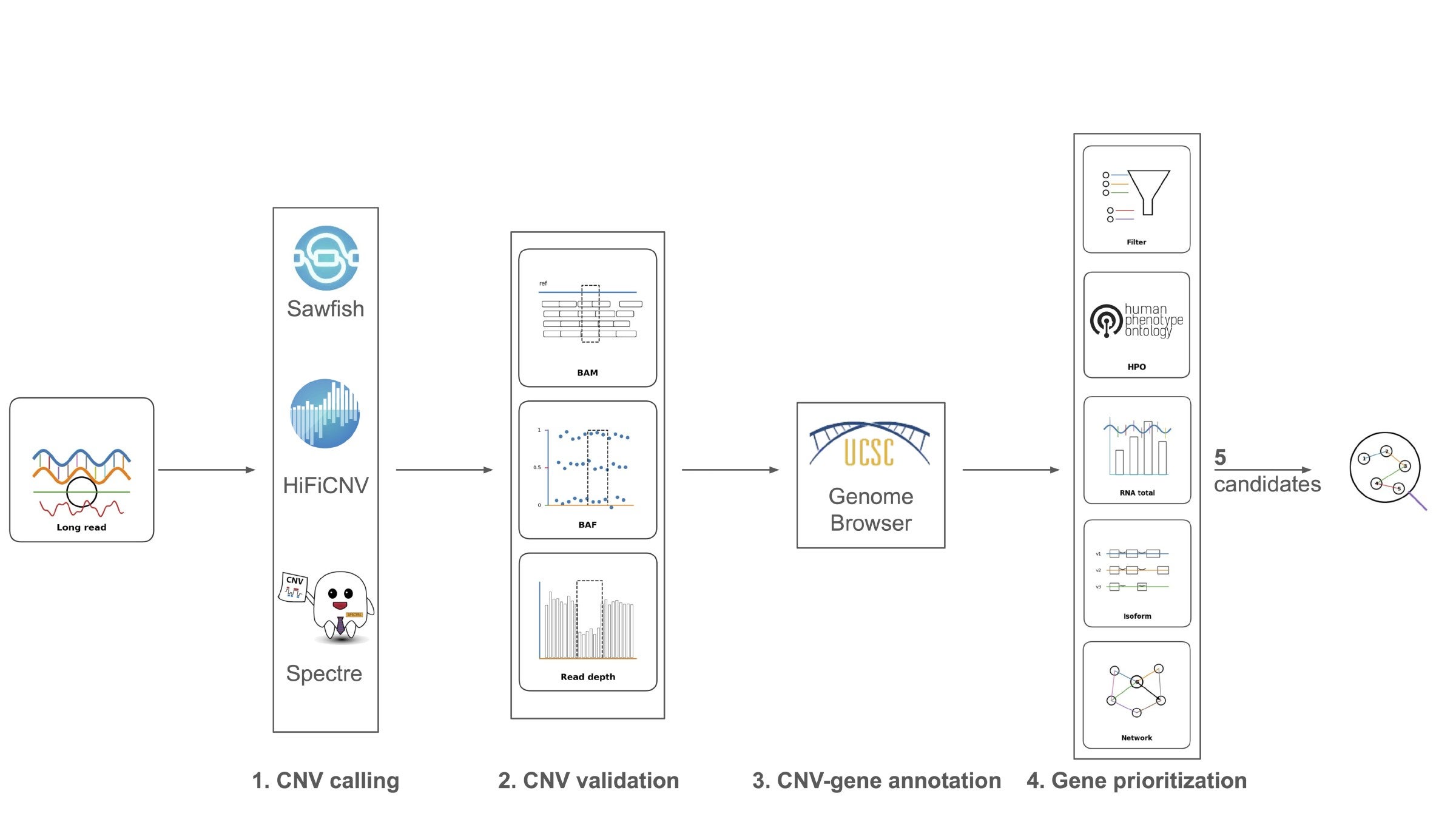
Figure 1. CNV-to-modifier-gene prioritization workflow. Long-read sequencing data were analyzed using three CNV callers, Sawfish, HiFiCNV, and Spectre, yielding 274 candidate CNVs. Candidate variants were manually reviewed using Binary Alignment Map (BAM) alignments, B-allele frequency (BAF), and read-depth evidence, resulting in 16 high-confidence CNVs. These CNVs were annotated against protein-coding genes using genome browser-based interval annotation, identifying 42 CNV-overlapped genes. Gene-level prioritization then integrated filtering criteria with four evidence layers: HPO-based phenotype matching, total RNA expression, isoform composition, and network proximity. This workflow produced five candidate modifier genes for mechanistic interpretation.
In Table 1, notation uses deletion (DEL), duplication (DUP), homozygous (hom), and heterozygous
(het).
| Tier | Gene | CNV locus (GRCh38) | Type (zyg.) | gnomAD–SV allele frequency | Evidence |
|---|---|---|---|---|---|
| 1 | LIMS3 | chr2:109.77– 110.10Mb (325kb) | DEL (hom) | rare | Expression reduced, consistent with the homozygous deletion (log2 fold-change −1*.*54). Two proteininteraction steps from ITGA3 (top 3% of genes). Paralogue of LIMS1/PINCH1, a focal-adhesion adaptor. |
| 2 | NOMO1 | chr16:14.895– 14.917Mb (22kb) | DUP (het) | rare | Expression strongly reduced despite the duplication (log2 fold-change −4*.82). Component of Nodal and transforming growth factor-β (TGFβ*) signalling. |
| 2 | EIF3C | chr16:28.65– 28.81Mb (161kb) | DEL (het) | 1.5% | Gene intolerant to loss of function; low-frequency deletion. |
| 2 | SESTD1 | chr2:179.202– 179.216Mb (14kb) | DEL (het) | 28.4% | Intronic deletion (coding sequence preserved); regulator of TRPC5 calciumchannel signalling. Common population polymorphism. |
| 2 2 | KCNE1 SMIM11 | chr21:34.38– 34.50Mb (116kb) | DEL (het) | 49.9% | Disease annotation limited to cardiac long-QT syndrome (no fibrosis link). Expression reduced; localises to focal adhesions. |
| 2 | SMIM34 | Small integral membrane protein; function uncharacterised. | |||
| 2 | SIK1 | chr21:43.37– 43.57Mb (206kb) | DEL (het) | 49.6% | Expression reduced; salt-inducible kinase 1, a TGF-β / SMAD2/3 target driving epithelial-to-mesenchymal transition. |
| 2 | HSF2BP | Meiotic recombination factor; no established fibrosis link. | |||
| 2 | ICOSLG | chr21:44.10– 44.25Mb (155kb) | DEL (het) | 47.6% | Immune co-stimulatory ligand; combined-immunodeficiency annotation matching the patient’s regulatory T-cell deficit. |
| 2 | GATD3 | Largest expression reduction in the dataset; mitochondrial deglycase linked to cellular senescence. | |||
| 2 | PWP2 | Expression reduced; ribosome biogenesis factor. | |||
| 3 | TRAPPC10 | chr21:44.10– 44.25Mb (155kb) | DEL (het) | 47.6% | Membrane-trafficking factor; canonical transcript remains dominant, indicating compensation. |
| 3p | RGPD5 | chr2:109.77– 110.10Mb (325kb) | DEL (hom) | rare | Discordant expression attributable to cross-mapping among RGPD-family paralogues; distant from ITGA3. |
Continued on next page
Tier | Gene | CNV locus (GRCh38) | Type (zyg.) | gnomAD–SV allele frequency | Evidence |
|---|---|---|---|---|---|
3p | TRIM73 | chr7:75.15– 75.42Mb (265kb) | DEL (hom) | rare | Expression reduced (log2 fold-change −1.22) but at very low read depth; cross-mapping among TRIM-family paralogues. |
– | EIF3CL | chr16:28.34– 28.46Mb (116kb) | DEL (het) | rare | Paralogue of EIF3C; signal arises from cross-mapping. |
– | 11 paralogue / segmentalduplication genes | various | – | GOLGA6L4, NPIPB6/8/9, TP53TG3C, TRIM49D2, TRIM64, FAM86B2, the eight-member SPDYE cluster, ARL17A/B, LRRC37A/A2; excluded for paralogue cross-mapping or absence from the Search Tool for the Retrieval of Interacting Genes/Proteins (STRING) network. | |
– | 15 no-evidence genes | various | – | Testis-restricted loci (the chrX NXF2/NXF2B and TCP11X1/X2 cluster) or genes with no expression in the available tissue and no curated annotation. | |
Table 1. Tiered candidate-gene prioritization and exclusion summary. Genes retained for modifier interpretation are grouped by evidence tier; excluded loci are summarized by the main reason for removal. Coordinates are GRCh38. Allele frequency is the maximum population frequency from gnomAD-SV; “rare” denotes a variant with no gnomAD-SV overlap or has allele frequency below 1%. Expression evidence is from long-read isoform sequencing, and log2 fold-change compares patient to control. DEL, deletion; DUP, duplication; hom, homozygous; het, heterozygous.
Five candidates show the strongest convergence across evidence layers
Five of the 13 retained genes LIMS3, NOMO1, ICOSLG, SIK1, and GATD3 were selected for detailed interpretation on the basis of either stronger convergence across the evidence layers or a particularly interpretable molecular mechanism connected to the patient’s disease context (Table 2). Each was supported by a different combination of layers. LIMS3 showed reduced RNA expression and the closest network proximity to ITGA3, connecting to the integrin focal-adhesion machinery through the parvin family (Figure 2). NOMO1 showed strongly reduced RNA expression despite a duplication, with a shift toward transcripts predicted to undergo nonsense-mediated decay, and joined the same network component through its paralogue NOMO3 (Figure 2). ICOSLG carried a truncating transcript and an immunodeficiency disease annotation, alongside the patient’s documented regulatory T-cell reduction. SIK1 showed reduced RNA expression. GATD3 showed the largest gene-dosage-loss signal in the dataset.
| Candidate | Proposed modifier axis | Supporting observations | Limitations |
|---|---|---|---|
| LIMS3 | Focal adhesion / integrin axis | Decreased RNA expression; close network proximity to ITGA3; focal-adhesion localization | Requires functional validation as a modifier of ITGA3-related disease |
| NOMO1 | Nodal / TGF-β signalling | Strongly decreased RNA expression despite a duplication; intolerance to loss of function | Effect inferred from regulatory/3′ untranslated-region (UTR) architecture rather than coding sequences |
| ICOSLG | Immune regulation / regulatory T-cell biology | Immune co-stimulatory ligand; immunodeficiency disease annotation; phenotype match to the patient’s documented regulatory T-cell deficit | Common CNV |
| SIK1 | TGF-β / SMAD epithelial-to-mesenchymal transition axis | Decreased RNA expression; TGF-β target driving epithelial-to-mesenchymal transition relevant to fibrosis | Common CNV |
| GATD3 | Mitochondrial dysfunction / senescence axis | Largest RNA expression reduction in the dataset; senescence biology plausibly linked to the salivary and renal phenotype | Common CNV |
Table 2. Summary of candidate modifier genes. Each candidate was prioritized through a different combination of molecular observation and biological plausibility in a single-proband (n = 1) analysis; the supporting observations are descriptive and hypothesis-generating. These genes are proposed as candidate modifiers that may help explain atypical features beyond the primary biallelic ITGA3 lesion. CNVs with allele frequency below 1% are considered common CNVs.
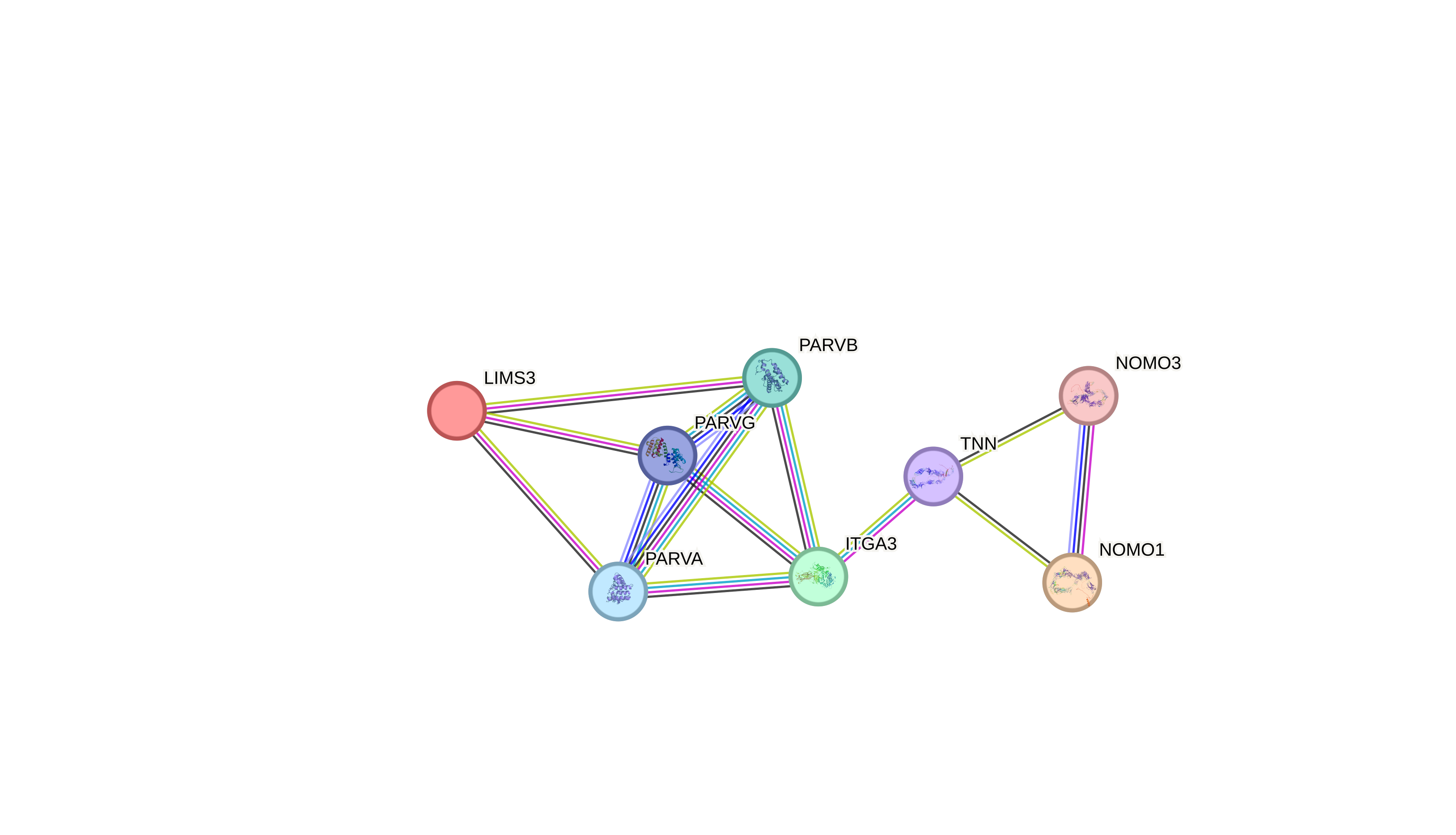
Figure 2. STRING PPI network linking ITGA3 to the candidate modifiers LIMS3 and NOMO1**.** The candidate modifiers were submitted to STRING (v12.0; minimum required interaction score ≥0.4), which added the five highest-confidence interactors needed to connect them. All eight proteins form a single connected component. LIMS3 connects to ITGA3 through the parvin family, recapitulating the IPP focal-adhesion complex, whereas NOMO1 associates with the component through its paralogue NOMO3. Edge colour denotes the source of supporting evidence: teal, curated databases; magenta, experimentally determined interactions; black, co-expression; lime, text mining; blue, gene co-occurrence; and light purple, protein homology. Edge thickness reflects the strength of the supporting data.
Discussion
This case is distinguished by an unusually atypical clinical course in a patient with biallelic loss of function in ITGA3. Biallelic ITGA3 disruption remains the primary molecular diagnosis and is sufficient to explain the core ILNEB disease mechanism, yet the patient’s survival into adulthood, progressive multi-organ fibrosis (pulmonary, renal, and salivary-gland), and immune dysregulation are not readily explained by the primary genotype alone. To investigate this gap, we applied a multi-layered, patient-partnered multi-omics workflow that consists of long-read CNV calling with manual validation, gene annotation, RNA expression and isoform analysis, phenotype matching, and protein–protein interaction (PPI) network evidence. This workflow asks whether additional copy-number variants might act as genetic modifiers by perturbing pathways related to the ITGA3associated cellular microenvironment, including basement-membrane integrity, focal adhesion, epithelial remodeling, fibrosis, immune regulation, and cellular stress responses. To our knowledge, this is the first application of a structured modifier-gene framework to an atypical ITGA3/ILNEB presentation.
Applying this framework yielded five candidate modifier genes whose biology plausibly intersects ITGA3-associated tissue injury. In nominating them, the analysis extends the two-hit, modifiergene paradigm to ILNEB. LIMS3 provides the most direct mechanistic link to the ITGA3 axis: it is a paralogue of LIMS1/PINCH1, the LIM-only adaptor that, with integrin-linked kinase and parvin, forms the IPP complex at integrin focal adhesions and stabilises the connection between integrins and the actin cytoskeleton32–33. Loss of PINCH1 in mouse epidermis causes detachment from the basement membrane, hair loss, and keratinocyte adhesion defects34, mirroring the basementmembrane fragility that underlies the ILNEB phenotype. Reduced LIMS3 dosage is therefore a coherent candidate amplifier of ITGA3-related adhesion failure. NOMO1 and SIK1 point toward dysregulated TGF-β-family signalling, a central driver of fibrosis. NOMO1, together with nicalin and TMEM147, forms an endoplasmic-reticulum complex that acts as a negative regulator of Nodal/Activin signalling within the TGF-β superfamily35–36; reduced functional NOMO1 would be predicted to relieve this restraint. SIK1 is a TGF-β–inducible kinase that provides negative feedback on the pathway, forming a complex with SMAD7 and SMURF2 to promote degradation of the type-I receptor and dampening TGF-β-driven transcription and epithelial-to-mesenchymal transition37–38. Its loss would similarly be expected to de-repress profibrotic signalling. ICOSLG connects the candidate landscape to immune regulation: it is the ligand for the inducible T-cell co-stimulator, and the ICOS/ICOSLG axis is required for the expansion, maintenance, and Foxp3 stability of regulatory T cells, while biallelic ICOSLG loss causes a combined immunodeficiency39–40, which directly matches the patient’s documented reduction in regulatory T cells. GATD3 is a mitochondrial-matrix deglycase that restricts the accumulation of advanced glycation end products, and its loss in mice causes advanced-glycation-end-product accumulation and altered mitochondrial dynamics41, providing a plausible route to the cellular stress and senescence biology associated with fibrotic tissue remodelling. Framing these variants as modifiers rather than incidental findings reframes how an atypical Mendelian presentation can be interpreted: each candidate maps onto a pathway already sensitized by the primary basement-membrane defect, generating specific, testable hypotheses about which biological axes shape disease expression.
These biological hypotheses carry clinical implications. For diagnosis, the primary genetic diagnosis is unchanged. The nominated candidates are not independent diagnoses but instead help contextualize an atypical expression of an established one. For prognosis, these modifiers offer a candidate explanation for the interindividual variability in severity, tissue involvement, and survival seen among patients sharing an ITGA3 diagnosis, and could ultimately better predict disease trajectory. For treatment, the implicated axes intersect pathways with existing or emerging interventions, including antifibrotic modulation of TGF-β signalling, support of regulatory T-cell function, and senescence-directed strategies such as senolytics.
These connections remain hypothesis-generating because of several limitations. The most fundamental is that the analysis rests on a single patient, so all modifier interpretations are hypothesisgenerating, cannot establish generalizable genotype–phenotype relationships, and carry no statistical support. Because DNA was available only for the proband, with no parental or family samples, we could not determine the inheritance or de novo status of candidate CNVs or formally separate private from inherited events. The RNA evidence derived from hair-follicle samples, which may not faithfully reflect expression in the tissues most relevant to the phenotype. CNV calling was performed in a single-sample, control-free setting. Without a matched control, distinguishing candidates from common CNV relied on public frequency resources and manual review. Some candidates additionally showed non-canonical dosage–expression relationships: increased expression following a deletion, or decreased expression following a duplication. This requires isoformand regulation-level interpretation rather than simple gene-dosage models. Finally, all proposed mechanisms remain functionally unvalidated.
These limitations warrant further further investigations. The next step would be to extend the analysis beyond one case and assembling a series of atypical or unusually severe ITGA3/ILNEB patients to test for recurrence, inheritance, and de novo status of modifiers. To address the tissue mismatch, expression and isoform evidence should be sought in disease-relevant tissues or cell types. The non-canonical dosage–expression relationships warrant targeted transcript-level validation, including reverse-transcription PCR (RT-PCR), allele-specific expression, and isoform quantification. Finally, the most actionable candidates should undergo orthogonal confirmation of CNV boundaries and direct functional testing, including perturbation and rescue in relevant cellular contexts, to establish whether they genuinely modify the consequences of the primary basement-membrane defect.
Conclusion
This patient-partnered analysis applies a staged, multi-evidence workflow to ask why this patient’s ITGA3-associated disease course is atypical. Long-read CNV calling, manual validation, gene annotation, RNA expression, isoform analysis, phenotype matching, and PPI network evidence together narrowed the search to five candidate modifier genes: LIMS3, NOMO1, SIK1, ICOSLG, and GATD3. These genes span focal adhesion, epithelial remodeling, immune regulation, membraneprotein biogenesis, and senescence. These candidates are not independent diagnoses, but they flag specific biological pathways that may shape how severely ITGA3-associated disease presents, turning one unusual case into a set of testable hypotheses. More broadly, the approach shows how patient-centered multi-omics analysis can carry rare-disease interpretation past a single causal variant, which is especially valuable for severe or atypical presentations, where a confirmed diagnosis still leaves much of the clinical picture unexplained.
Acknowledgements
We are deeply grateful to the patient, whose willingness to participate and to share genomic, transcriptomic, and clinical data made this patient-partnered study possible. We thank Dr. Tushar Desai for clinical care and clinical characterization of the proband. We thank German Beldorati Stark for data processing and bioinformatics support that prepared the sequencing data for downstream analysis. We also acknowledge the Allman Institute for its partnership in this patient-partnered case study.
Funding
This work was supported by the Research to the People.
Be the first to comment on this publication.