Purpose
A patient with biallelic ITGA3 pathogenic variants presented with severe lung fibrosis, along with renal involvement and mild skin abnormalities. The molecular mechanisms underlying this disease at the spatial and cellular levels remain poorly understood. Spatial transcriptomics (Xenium) was performed on lung tissues from this patient at two time points (2017 and 2022). Cell type composition and global transcriptional structure were stable across time. In contrast, transcriptional comparison between histologically normal and fibrotic regions revealed extracellular matrix (ECM) activation in fibrotic regions, while ITGA3 was differentially expressed but showed a non–region-specific distribution. These findings support a model in which epithelial dysfunction drives multicellular ECM remodeling in lung fibrosis and provide insight into potential therapeutic strategies targeting core fibrotic pathways.
Introduction
Pulmonary fibrosis encompasses a group of chronic, progressive lung disorders characterized by excessive extracellular matrix deposition and irreversible distortion of normal lung architecture, ultimately leading to respiratory failure. Although numerous risk factors for pulmonary fibrosis have been identified, the cellular and molecular mechanisms underlying fibrosis driven by rare pathogenic variants remain incompletely understood.
Integrin α3, encoded by ITGA3, is broadly expressed in epithelial tissues, including airway and alveolar epithelium, as well as in skin and kidney. Biallelic pathogenic variants in ITGA3 cause a multisystem disorder known as ILNEB syndrome (interstitial lung disease, nephrotic syndrome, and epidermolysis bullosa)1. Experimental studies have shown that α3β1 integrin plays a critical role in epithelial adhesion to the basement membrane and in maintaining epithelial integrity2–3. Disruption of integrin-mediated adhesion has been linked to epithelial injury, altered cell–matrix signaling, and activation of fibrogenic pathways, including transforming growth factor–β (TGF-β) signaling and epithelial–mesenchymal transition (EMT)4. However, the contribution of ITGA3 dysfunction to human lung fibrosis, particularly in the context of spatial tissue organization, remains poorly defined.
The patient studied here carries biallelic pathogenic variants in ITGA3, including a splice-site loss-of-function mutation and a non-conservative missense variant (Thr658Arg), and presents with advanced lung disease characterized by both airway-predominant fibrosis and interstitial fibrosis. Histopathology demonstrates epithelial sloughing and marked collagen deposition, consistent with impaired epithelial adhesion and progressive fibrotic remodeling. This combination of epithelial instability and extensive ECM accumulation provides a unique opportunity to examine how epithelial defects translate into multicellular fibrotic programs in human lung tissue.
Recent advances in spatial transcriptomics, including Xenium, enable high-resolution mapping of gene expression within intact tissue architecture, allowing direct integration of cellular identity, spatial context, and transcriptional state. Such approaches are particularly valuable in rare, genetically defined cases, where patient-specific molecular profiling can provide mechanistic insights and inform precision medicine strategies. In this study, spatial RNA profiling was applied to lung wedge resections obtained in 2017 and 2022 from this patient with ITGA3-associated pulmonary fibrosis. The analysis focuses on identifying cell type–specific and region-specific transcriptional programs, with particular emphasis on pathways associated with ECM remodeling and their relationship to underlying epithelial dysfunction.
Results
No major differences in cell distribution or composition between 2017 and 2022
Raw Xenium data from 2017 and 2022 lung wedge resection samples showed comparable distributions of detected features and transcript counts per cell (Figure 1A). Quality control filtering removed a small fraction of cells in both datasets, including cells without detected transcripts (2017: 0.24%; 2022: 0.27%), cells with high negative control counts (2017: 0.00%; 2022: 0.00%), and cells exceeding 5× median absolute deviation (MAD) (2017: 0.92%; 2022: 1.03%). After filtering, 632,371 cells from 2017 and 728,081 cells from 2022 were retained, with similar distributions of feature counts and transcript counts (Figure 1B).
Cell type annotation revealed highly comparable cellular compositions between the two time points (Figure 1C). Major epithelial, mesenchymal, immune, and endothelial populations were present in both datasets at similar proportions, indicating stability of overall cell type composition over time. No substantial enrichment or depletion of specific cell populations was observed.
Dimensionality reduction using UMAP demonstrated strong overlap between cells from 2017 and 2022, with no clear segregation by time point (Figure 1D). Consistently, annotated cell types occupied similar regions in the UMAP space in both datasets (Figure 1E), indicating that transcriptionally defined cell identities were preserved across time. Together, these findings indicate that both global cell distribution and cell type composition remained stable between 2017 and 2022 in this patient.

Figure 1. Stable cell composition and distribution across 2017 and 2022 lung samples.**(A) Distribution of detected gene features (nFeature_Xenium) and transcript counts (nCount_Xenium) per cell in raw Xenium data from 2017 and 2022 samples.(B) Distributions of gene features and transcript counts after quality control filtering.(C) Proportions of major cell types across epithelial, mesenchymal, immune, and endothelial compartments in 2017 and 2022, showing similar cell type composition between time points.(D) UMAP projection of all cells from both datasets, colored by sample year, demonstrating strong overlap between 2017 and 2022 cells.(E) UMAP projections of 2017 and 2022 datasets with cell type annotations, showing that corresponding cell types occupy similar regions in low-dimensional space across time.
Extracellular matrix–associated transcriptional programs in fibrotic areas
Given the absence of major temporal differences in cell composition, analyses were focused on spatial differences between histologically normal-appearing and fibrotic regions. Distinct regions were annotated on tissue sections, with fibrotic areas and normal areas clearly delineated (Figure 2A).
Cell type–resolved quantification within these regions showed that multiple epithelial, mesenchymal, immune, and endothelial populations were present in both compartments (Figure 2B). To ensure robust downstream comparisons, only cell types with sufficient representation and without extreme imbalance in cell number between regions were retained for differential gene expression analysis.
Differential expression analysis within each retained cell type identified variable numbers of differentially expressed genes (DEGs) (Figure 2C). Mesenchymal populations, particularly myofibroblasts and smooth muscle cells, exhibited the highest numbers of DEGs, while epithelial and immune cell types showed more moderate changes. This pattern suggests that mesenchymal compartments undergo more pronounced transcriptional alterations in fibrotic regions.
Functional enrichment analysis showed that differential gene expression converged on a limited number of major functional categories (Figure 2D). The most prominent category was extracellular matrix (ECM) (extracellular structure organization, collagen fibril organization, external encapsulating structure organization, and cell–matrix adhesion). These terms were consistently enriched across multiple cell types. Vascular remodeling (regulation of angiogenesis, vasculature development, endothelial development) and immune and injury-related processes (leukocyte migration, mononuclear cell migration, wound healing, phagocytosis) were less consistent across cell types. Other pathways, including epithelial/developmental processes (lung development, respiratory tube development, morphogenesis of a branching epithelium) and fibrosis-related signaling (transforming growth factor beta production, integrin-mediated signaling), were detected in a subset of cell types.
Individual ECM-associated genes showed widespread regulation in fibrotic regions across several cell types (Figure 2E). Genes encoding structural ECM components (e.g., collagens), matrix-modifying enzymes (e.g., MMPs), and ECM-associated regulators (e.g., POSTN, THBS1) showed increased expression and statistical significance in multiple lineages, particularly within mesenchymal and endothelial cells. Notably, several of these genes were also upregulated in epithelial and immune populations, suggesting that ECM remodeling is not restricted to classical stromal compartments but represents a coordinated, multicellular response in fibrotic tissue.
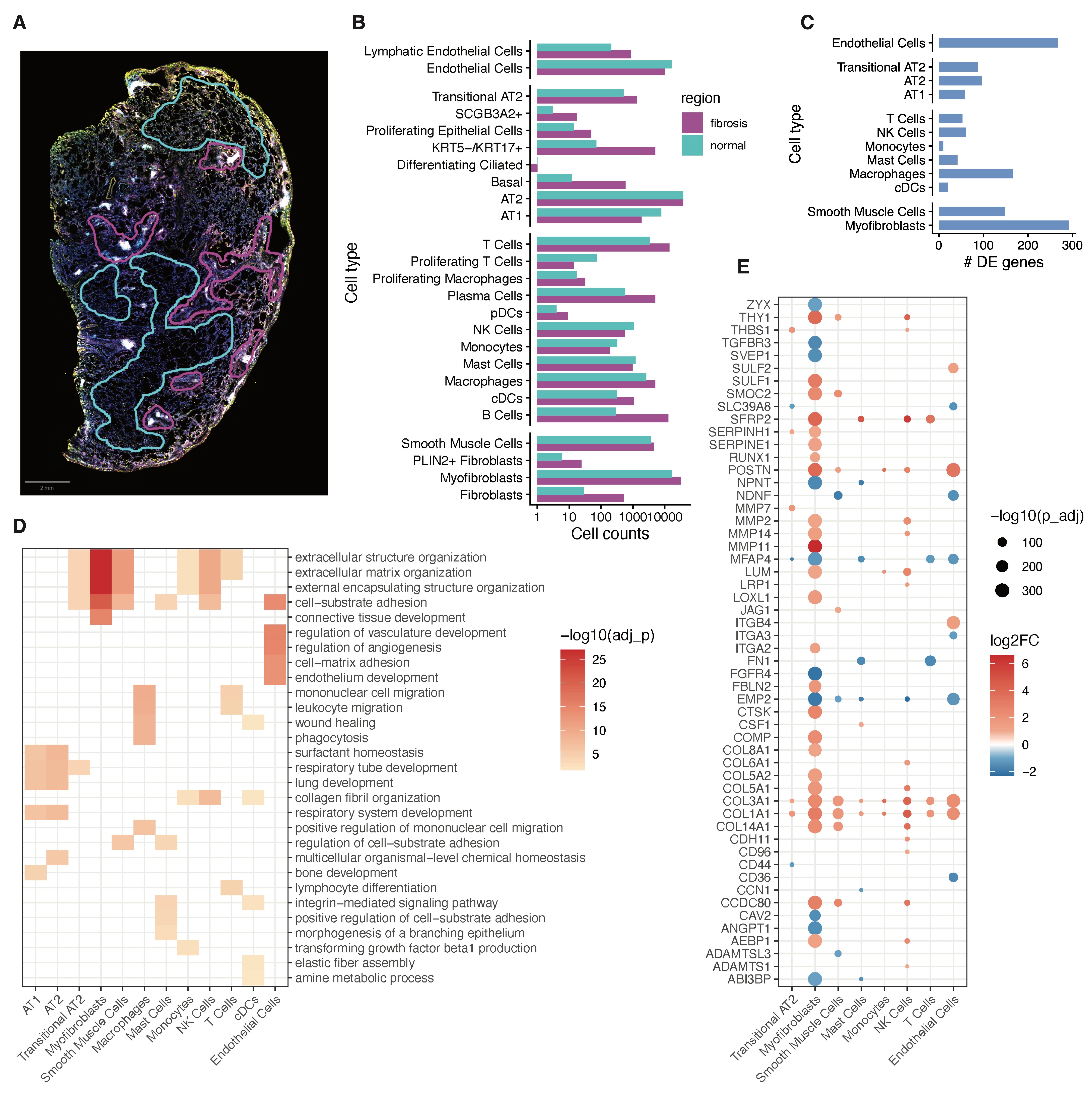
Figure 2. Extracellular matrix remodeling in fibrotic regions.**(A) Representative tissue section with annotated regions corresponding to histologically normal-appearing areas (blue) and fibrotic areas (purple).(B) Cell type–specific cell counts in normal and fibrotic regions.(C) Number of differentially expressed genes (DEGs) identified within each cell type between fibrotic and normal regions.(D) Heatmap of functional enrichment analysis for DEGs across cell types, showing significant enrichment of extracellular matrix–related pathways and associated biological processes.(E) Dot plot showing fold change and statistical significance of ECM-associated genes across cell types, highlighting widespread upregulation of matrix-related genes in fibrotic regions.
ECM gene expression is enriched in fibrotic regions
An ECM gene signature score projected onto tissue sections showed higher values in fibrotic regions compared to histologically normal-appearing areas, indicating spatial enrichment of ECM-related transcriptional activity (Figure 3A). In contrast, ITGA3 expression appeared diffuse across the tissue, without clear spatial enrichment in fibrotic regions (Figure 3B) despite its differential expression in cell type–resolved analysis.
Additional ECM-associated genes (e.g., COL1A1, POSTN, MMP14, and SFRP2) showed higher expression in fibrotic regions, with visibly stronger signal intensity compared to normal areas (Figure 3C–F). These patterns were consistent with the enrichment of ECM-related pathways identified in differential expression and functional analyses, supporting localized activation of matrix remodeling programs in fibrotic tissue.
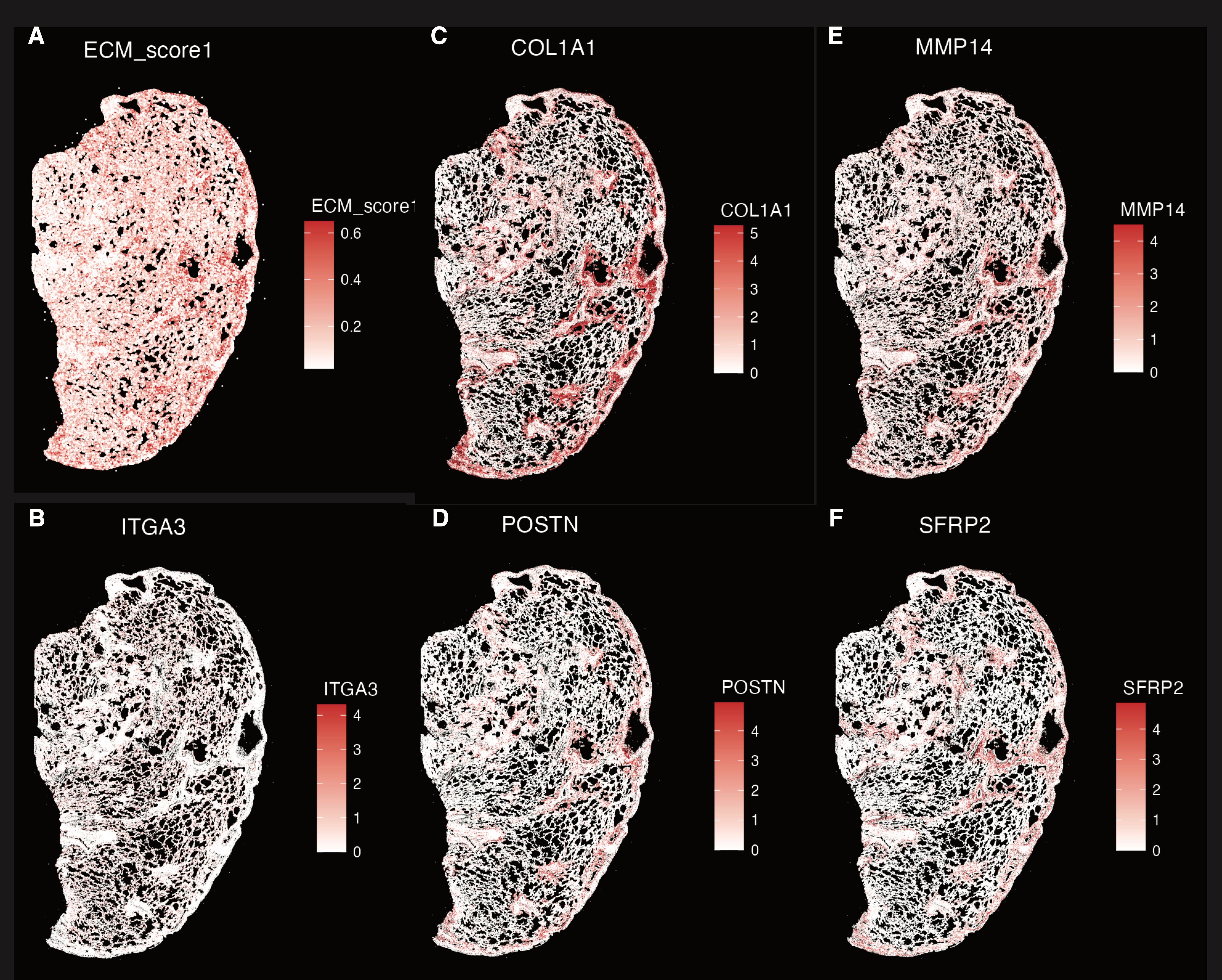
Figure 3. Localized activation of ECM-related gene expression in fibrotic regions.**(A) Spatial distribution of ECM gene signature scores across the tissue section, showing higher scores in fibrotic regions.(B) Spatial expression of ITGA3, showing diffuse distribution without clear enrichment in fibrotic regions.(C–F) Spatial expression of representative ECM-associated genes (COL1A1, POSTN, MMP14, SFRP2), showing increased expression in fibrotic regions compared to normal areas.
Discussion
This study provides a spatially resolved view of advanced lung fibrosis in a patient with biallelic ITGA3 pathogenic variants, integrating cell type composition, regional transcriptional changes, and tissue architecture. Despite the long disease course and interval between sampling (2017 vs 2022), cell type composition and global transcriptional structure remained stable, indicating that disease progression is not driven by major shifts in cellular populations. Instead, spatial context emerged as the dominant source of variation, with fibrotic regions showing consistent transcriptional differences across multiple cell types.
Across epithelial, mesenchymal, endothelial, and immune compartments, extracellular matrix (ECM)–related programs represented the most prominent and consistent signal in fibrotic regions. Enrichment of pathways related to collagen organization, matrix assembly, and cell–matrix interaction, together with increased expression of structural ECM genes and matrix-remodeling enzymes, supports a model of coordinated, multicellular activation of fibrogenic processes. While mesenchymal cells showed the strongest transcriptional changes, ECM-associated gene upregulation was also observed in non-mesenchymal populations, indicating that matrix remodeling in advanced fibrosis involves broad participation across cell types rather than being restricted to classical fibroblast lineages.
Although ITGA3 was identified as differentially expressed in cell type–resolved analyses, its spatial expression was not regionally enriched, suggesting that its role is not confined to fibrotic niches but may instead reflect a global alteration in epithelial or tissue-level biology. ITGA3 encodes integrin α3, a key component of α3β1 integrin complexes that mediate epithelial adhesion to the basement membrane and regulate signaling pathways linked to cell survival, polarity, and matrix interaction. Prior studies have shown that disruption of integrin-mediated adhesion can impair epithelial integrity and promote fibrotic remodeling through enhanced susceptibility to injury and dysregulated TGF-β and integrin signaling. In this context, the diffuse expression pattern observed here is consistent with a model in which ITGA3 dysfunction contributes to baseline epithelial instability, which in turn facilitates chronic injury and secondary activation of ECM-producing programs across multiple cell types.
These findings have potential therapeutic implications. The dominance of ECM-related pathways across cell types supports targeting core fibrotic processes, such as collagen production, matrix crosslinking, or integrin-mediated signaling, rather than focusing on individual cell populations. In addition, the lack of spatial restriction of ITGA3 expression suggests that interventions aimed at restoring epithelial adhesion or modulating integrin signaling may need to act broadly across the tissue. Given the patient’s clinical course, characterized by exacerbations following viral infections, strategies that reduce injury-induced amplification of fibrotic responses may also be relevant. Overall, this study supports a model in which genetically driven epithelial dysfunction establishes a permissive environment for widespread, multicellular ECM remodeling, ultimately leading to progressive lung fibrosis.
Methods
Spatial transcriptomics data processing
Spatial transcriptomic data were generated from two FFPE lung tissue sections (2017 and 2022) using the 10x Genomics Xenium platform. Raw data were imported into R using the Seurat package (v5) via LoadXenium. Separate Seurat objects were created for each time point and annotated with sample metadata.
Quality control
Quality control was performed independently for each dataset. Cells were excluded if they met any of the following criteria: (1) zero detected transcripts (nCount_Xenium = 0); (2) high background signal, defined as a negative control fraction ≥ 0.1 (nCount_BlankCodeword / nCount_Xenium); or (3) abnormally high transcript counts, defined as greater than 5 median absolute deviations (MAD) above the median nCount_Xenium. Distributions of gene counts (nFeature_Xenium) and transcript counts (nCount_Xenium) were visualized using VlnPlot.
Dimensionality reduction and cell type annotation
A subset of 30,000 cells per sample was used to construct a reference dataset. Data were normalized (NormalizeData), variable features identified (FindVariableFeatures, n = 3000), scaled (ScaleData), and subjected to principal component analysis (RunPCA). Uniform manifold approximation and projection (UMAP) was computed using RunUMAP, followed by graph-based clustering (FindNeighbors, FindClusters, resolution = 0.3).
Cell type annotation was performed using label transfer from a published human lung reference dataset5. Transfer anchors were identified using FindTransferAnchors, and cell types were assigned using MapQuery. The annotation model was subsequently applied to the full datasets using a chunk-based approach to accommodate large cell numbers.
Cell type composition analysis
Cell type proportions were calculated from combined metadata across time points using contingency tables and normalized to percentages. Cell types with low abundance (<1%) were excluded from visualization. Results were visualized using ggplot2.
Region annotation and cell filtering for differential expression
Regions of interest (fibrotic vs histologically normal) were defined based on external annotation and mapped to individual cells. For differential expression analysis, cell types were retained if they met the following criteria: (1) ≥50 total cells; (2) ≥10 cells in each region; and (3) balanced representation between regions (fibrotic cell fraction between 0.1 and 0.9).
Differential expression analysis
Within each retained cell type, differential gene expression between fibrotic and normal regions was performed using FindMarkers (Seurat), with thresholds of minimum expression fraction ≥0.1 (min.pct) and log2 fold change ≥0.25. Genes with adjusted p-value <0.05 were considered significant.
Functional enrichment analysis
Gene Ontology (GO) enrichment analysis was performed using clusterProfiler (enrichGO, ontology = “BP”) with gene ID conversion via bitr (org.Hs.eg.db). Enriched terms with adjusted p-value <0.05 were retained. Top pathways per cell type were visualized as a heatmap based on −log10(adjusted p-values).
ECM gene set analysis and spatial visualization
Extracellular matrix (ECM)–related genes were defined from enriched GO terms, including extracellular structure organization and cell–matrix adhesion pathways. An ECM gene signature score was computed using AddModuleScore and visualized spatially using ImageFeaturePlot.
ECM-related differentially expressed genes were further extracted across selected cell types, filtered by absolute log2 fold change >1, and visualized using dot plots to display effect size and statistical significance.
Be the first to comment on this publication.