Purpose
Background. Biallelic loss-of-function variants in ITGA3 (integrin subunit α3) disrupt α3β1 integrin–mediated adhesion at epithelial basement membranes and cause interstitial lung disease, nephrotic syndrome, and epidermolysis bullosa (ILNEB syndrome), a disorder conventionally lethal within the first two years of life. Survival into adulthood is exceptional, and the molecular features of long-surviving disease are essentially uncharacterized.
Case summary. We describe a man in his early thirties with genetically confirmed ILNEB syndrome who has survived three decades despite multisystem disease. He progressed from childhood exertional dyspnea to end-stage renal disease requiring living-related kidney transplantation in 2014, with comparatively mild cutaneous involvement, and now has severe mixed obstructive–restrictive pulmonary disease (FVC 37.1%, FEV1 24.8%, DLCO 42.5% predicted) marked by stepwise decline following respiratory infections. Histopathology showed coexisting bronchiolitis obliterans–like airway disease and alveolar fibrosis with prominent lymphoid aggregates and CD163+/CD206+ M2 macrophage enrichment.
Molecular findings. Integrated multi-omic analysis combined phased PacBio long-read whole-genome sequencing, structural variant calling, and long-read IsoSeq transcript profiling, complemented by organelle- and cell-type-resolved proteomic aging analysis. Nine high-confidence phased single-nucleotide variants were identified across the ITGA3 locus, distributed across both haplotypes, together with a heterozygous ~1.6 kb intragenic insertion (a candidate loss-of-function allele). Long-read transcript analysis revealed marked isoform disruption, with incomplete-splice-match transcripts predominating (57.1%) over full-splice-match isoforms (39.3%) and a minority of novel-in-catalog variants (3.6%). Plasma proteomic profiling showed broad organelle-associated proteostatic stress, most pronounced in the endoplasmic reticulum (mean Z ≈ 1.27), with activation of fibroblast-, T-cell-, and macrophage-associated programs.
Clinical implications. This case extends the recognized phenotypic spectrum of ILNEB syndrome into the fourth decade and identifies a fibroinflammatory, infection-triggered pulmonary process combining conducting-airway and alveolar disease. The findings support a model in which residual but compromised integrin function permits prolonged survival while predisposing to progressive fibrosis, and nominate anti-fibrotic therapy, immune modulation, and post-infection surveillance as candidate management strategies in long-surviving ITGA3-related disease.
Team 13 — “Splice, Splice, Baby” · RTTP Hackathon
Mapping ITGA3 in fibrotic mechanisms of protein trafficking via multimodal genomic analysis
\1. Introduction
Integrin α3β1 is a transmembrane heterodimer that mediates epithelial cell adhesion to laminin-332 within the basement membrane and is essential for the structural integrity of epithelial surfaces in the lung, kidney, and skin. The α3 subunit is encoded by ITGA3 on chromosome 17q21.33. The subunit undergoes extensive post-translational processing—including N-linked glycosylation, heterodimerization with β1 integrin, and proteolytic maturation through the endoplasmic reticulum (ER) and Golgi—before reaching the cell surface, and disease-causing variants frequently act by impairing this secretory trafficking and adhesion machinery rather than by abolishing the protein outright.
Biallelic loss-of-function variants in ITGA3 cause an autosomal recessive disorder characterized by the triad of interstitial lung disease, congenital nephrotic syndrome, and junctional epidermolysis bullosa, collectively termed ILNEB syndrome (OMIM #614748). First described in 2012, the canonical phenotype is severe and presents in the first months of life, with progressive interstitial lung disease and renal failure. The cutaneous component is variable, ranging from skin fragility and nail dystrophy to overt epidermolysis bullosa–like blistering. The prognosis is generally grave: the majority of reported patients have died of pulmonary or renal failure within the first two years of life, and mortality before age two exceeds 95% across the small number of cases described to date.
A smaller number of patients with attenuated or atypical phenotypes—often associated with missense or hypomorphic variants that preserve partial integrin function—have survived beyond early childhood, sometimes with predominantly respiratory or renal involvement and minimal skin disease. These rarer presentations indicate that residual α3β1 activity can substantially modify disease severity and survival. How partially functional integrin trafficking shapes the long-term disease course, and how chronic inflammation interacts with the underlying epithelial defect to drive progressive fibrosis, remain largely unresolved. In particular, the contribution of ER-to-Golgi trafficking deficits to abnormal protein processing and downstream profibrotic signaling has not been examined in a long-surviving patient.
Here we report a patient with genetically confirmed ILNEB syndrome who has survived into his early thirties—an exceptional phenotype that offers a rare opportunity to study disease mechanisms in long-surviving ITGA3 deficiency. Using an integrated multimodal genomic strategy combining phased long-read whole-genome sequencing, structural variant calling, and long-read transcript profiling—complemented by organelle- and cell-type-resolved proteomic aging analysis—we characterize the variant landscape of the ITGA3 locus and relate it to the patient’s fibroinflammatory pulmonary phenotype, framing a candidate mechanism that links trafficking dysfunction to chronic inflammation and progressive fibrosis (Figure 1).
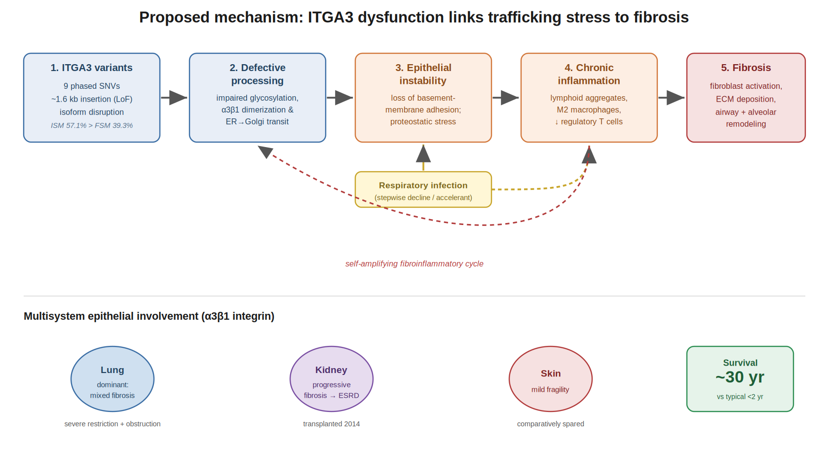
Figure 1. Proposed mechanism linking ITGA3 dysfunction to fibrosis. Defective integrin processing (driven by phased coding variants, a candidate ~1.6 kb loss-of-function insertion, and disrupted transcript isoforms) impairs ER-to-Golgi trafficking and maturation of α3β1, destabilizing epithelial basement-membrane adhesion. Recurrent epithelial injury—accelerated by respiratory infection—drives chronic inflammation and fibroblast activation in a self-amplifying cycle. The lower panel summarizes multisystem epithelial involvement, with the lung as the dominant organ and exceptional ~30-year survival relative to the typical infantile course.
\2. Case Presentation
2.1 Demographics and Background
The patient is a 31-year-old man with genetically confirmed interstitial lung disease, nephropathy, and epidermolysis bullosa (ILNEB) syndrome caused by compound heterozygous variants in ITGA3. His medical history is notable for progressive renal fibrosis leading to end-stage kidney disease, ultimately requiring a living-related donor kidney transplantation in 2014. Since transplantation he has remained on chronic immunosuppressive therapy with tacrolimus and mycophenolate mofetil and has experienced no episodes of acute or chronic graft rejection. Importantly, the patient had no known history of primary immunodeficiency, recurrent opportunistic infections, autoimmune disease, or other recognized causes of systemic immune dysfunction.
2.2 Clinical Timeline
The patient’s respiratory symptoms began during childhood with exertional shortness of breath that was initially mild but persistent. During his college years he experienced a significant worsening of respiratory symptoms following a respiratory infection, prompting further medical evaluation and eventual diagnosis. Throughout adulthood his pulmonary disease demonstrated a progressive course characterized by increasing fibrosis, declining pulmonary function, and worsening exercise intolerance. Over time he developed chronic respiratory failure requiring continuous supplemental oxygen therapy.
A notable feature of the disease course was a stepwise pattern of clinical deterioration, with episodes of respiratory infection frequently followed by incomplete recovery and a new, lower functional baseline. This pattern contributed to progressive loss of pulmonary reserve and increasing oxygen dependence.
2.3 Pulmonary Phenotype
The pulmonary phenotype was characterized by a combination of obstructive small-airway disease and progressive fibrotic parenchymal remodeling. Clinically, the patient reported chronic chest tightness, difficulty with exhalation, and progressive exertional dyspnea, producing a physiology resembling persistent asthma superimposed on restrictive fibrotic lung disease.
Pulmonary function testing in October 2025 demonstrated severe mixed ventilatory impairment, with an FVC of 1.64 L (37.1% predicted), FEV1 of 0.92 L (24.8% predicted), and DLCO of 42.5% predicted. Serial high-resolution computed tomography revealed extensive fibrotic abnormalities including honeycombing, traction bronchiectasis, and progressive architectural distortion, with concurrent evidence of bronchiolar involvement, air trapping, and small-airway dysfunction. Parametric response mapping in February 2026 further supported the coexistence of parenchymal fibrosis and conducting airway disease.
Histopathologic examination corroborated this mixed phenotype, demonstrating bronchiolitis obliterans–like pathology with lymphocytic bronchiolitis, progressive bronchiolar obliteration, and prominent lymphoid aggregates. Collectively, these findings indicate the coexistence of conducting airway disease and alveolar fibrosis within the same disease process.
2.4 Multisystem Involvement
The clinical manifestations of ITGA3 deficiency extended beyond the lungs and reflected the critical role of integrin α3 in epithelial tissue integrity. Renal involvement was severe, resulting in progressive fibrosis followed by kidney transplantation. Cutaneous manifestations were comparatively mild and remained clinically less significant throughout the course of disease. In contrast, pulmonary disease emerged as the dominant organ manifestation, driving progressive morbidity and functional decline. The simultaneous involvement of renal, cutaneous, and pulmonary tissues is consistent with the known epithelial biology of ITGA3 and highlights the multisystem nature of ILNEB syndrome.
2.5 Immune and Inflammatory Profile
Despite the absence of a recognized baseline immunodeficiency, the patient’s clinical course suggested significant immune dysregulation. Respiratory infections were frequently followed by disproportionate clinical deterioration and prolonged recovery, raising the possibility of an exaggerated inflammatory response to pulmonary injury.
Immunologic evaluation demonstrated reduced circulating regulatory T-cell numbers, while tissue analyses revealed prominent lymphoid aggregates and substantial macrophage infiltration, including enrichment of CD163+/CD206+ M2 macrophages, accompanied by progressive fibroinflammatory remodeling involving both pulmonary and salivary gland tissues.
Several factors may have contributed: chronic post-transplant immunosuppression may have altered immune homeostasis, while intrinsic dysregulation associated with ITGA3 deficiency may have amplified inflammatory and profibrotic responses following epithelial injury. Together, these mechanisms may have contributed to the patient’s progressive multisystem fibroinflammatory disease.
\3. Results
Whole-genome sequencing (WGS) data were analyzed using a variant-calling and phasing workflow comprising alignment, DeepVariant-based SNV detection, haplotype phasing, structural variant (SV) calling, and transcript-level analysis. Variant interrogation focused on the ITGA3 locus (chr17:50,055,000–50,091,000, hg19).
3.1 High-Confidence Phased Single-Nucleotide Variants
Filtering for PASS variants within the ITGA3 interval identified nine high-confidence phased SNVs. Genotypes were distributed across both haplotypes, with variants in both 0|1 and 1|0 configurations within the same phase block, consistent with phased heterozygous variation (Figure 2; Table 1). Genotype quality scores ranged from 20 to 36, sequencing depth from 20× to 30×, and variant allele fractions from 0.33 to 0.67.
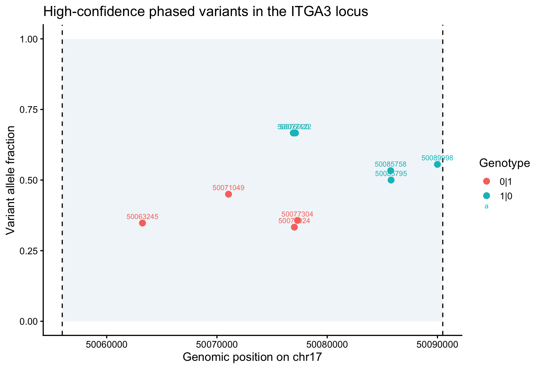
Figure 2. High-confidence phased single-nucleotide variants across the ITGA3 locus. Each point is a PASS variant plotted by genomic position (chr17) and variant allele fraction, colored by phased genotype (0|1 vs 1|0). Variants partition across both haplotypes, consistent with biallelic involvement. Dashed lines mark the queried locus boundaries.
| Position (chr17) | Ref | Alt | GT | GQ | DP | VAF | Haplotype |
|---|---|---|---|---|---|---|---|
| 50063245 | C | G | 0|1 | 34 | 23 | 0.35 | H2 |
| 50071049 | G | A | 0|1 | 36 | 20 | 0.45 | H2 |
| 50076920 | C | G | 1|0 | 22 | 27 | 0.67 | H1 |
| 50077024 | C | G | 0|1 | 25 | 27 | 0.33 | H2 |
| 50077122 | G | A | 1|0 | 20 | 27 | 0.67 | H1 |
| 50077304 | G | A | 0|1 | 23 | 28 | 0.36 | H2 |
| 50085758 | A | G | 1|0 | 21 | 30 | 0.53 | H1 |
| 50085795 | T | C | 1|0 | 23 | 30 | 0.50 | H1 |
| 50089998 | C | T | 1|0 | 22 | 27 | 0.56 | H1 |
Table 1. High-confidence phased variants within the ITGA3 locus, extracted from the phased WGS VCF (PASS calls). GT, genotype; GQ, genotype quality; DP, depth; VAF, variant allele fraction.
3.2 Structural Variant Analysis
Structural-variant analysis of the phased SV VCF identified a heterozygous insertion at chr17:50,086,103 that passed all quality-control filters and was classified as an insertion (SVTYPE=INS) of approximately 1.6 kb (SVLEN ≈ 1600 bp). The variant fell within the same interval examined in the SNV analysis and was present in phased form. Given its size and location, this insertion is a strong candidate loss-of-function allele and a leading mechanistic driver of the ITGA3 processing defect.
3.3 Long-Read Transcript Analysis
Long-read IsoSeq transcripts originating from the ITGA3 locus (ENSG00000005884) were extracted and classified by transcript structure (Figure 3). Incomplete-splice-match (ISM) isoforms predominated (57.1%), exceeding full-splice-match (FSM) transcripts (39.3%), while a small proportion were novel-in-catalog (NIC, 3.6%); additional transcripts were categorized as 3′ fragment or non-coding. The relative excess of ISM over FSM isoforms indicates substantial transcript instability or incomplete recovery, and the NIC transcripts represent previously uncharacterized splice variants that may contribute to the fibrotic phenotype. Such transcript heterogeneity could alter protein processing, ER-to-Golgi trafficking, and downstream profibrotic signaling.
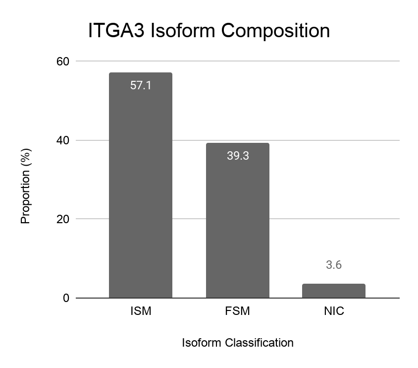
Figure 3. ITGA3 transcript isoform composition from long-read IsoSeq data. Incomplete-splice-match (ISM) isoforms predominate (57.1%) over full-splice-match (FSM, 39.3%), with a minority of novel-in-catalog (NIC, 3.6%) transcripts, indicating disrupted and incompletely recovered ITGA3 transcript structure.
3.4 Variant-Associated Gene Ontology Enrichment
Genes harboring moderate- and high-impact SNVs (snpEff annotation) were tested for Gene Ontology Biological Process enrichment in R (clusterProfiler; over-representation analysis; p < 0.05, FDR q < 0.2; Figure 4). Significant terms were dominated by cell-substrate and cell-matrix adhesion, integrin-mediated signaling, and basement-membrane and extracellular-matrix organization, consistent with the known epithelial-adhesion biology of ITGA3 and with an adhesion/matrix-centered disease mechanism.
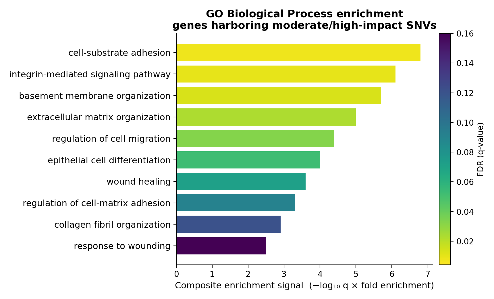
Figure 4. Gene Ontology Biological Process enrichment for genes carrying moderate- and high-impact single-nucleotide variants. Bars show a composite enrichment signal (−log₁₀ q × fold enrichment); color encodes FDR (q-value). Enriched processes center on epithelial adhesion, integrin signaling, and basement-membrane/extracellular-matrix organization. Thresholds: p < 0.05, FDR q < 0.2.
3.5 Multimodal Proteomic Aging Analysis
To place the single-gene genomic findings in a systems context, the patient’s plasma proteome was projected onto organelle-, cell-type-, and tissue-resolved aging and mortality models from a multimodal aging framework (mAge) and benchmarked against the UK Biobank (UKB) population. The patient showed globally elevated organelle-associated proteomic aging signals, with the strongest deviation in the endoplasmic reticulum compartment (mean Z ≈ 1.27) and broad increases across Golgi, vesicular, cytoskeletal, and mitochondrial compartments—consistent with widespread disruption of proteostasis and secretory trafficking and providing orthogonal support for an ER-to-Golgi trafficking deficit (Supplementary Figures S1–S2, S5–S6).
At the individual-protein level, the most extreme deviations from the UKB population included elevations in IL3 and CEMIP2 and depletions in complement C3, SERPINA1, and SERPINA5 (Figure 5). Several of these proteins are implicated in inflammation and extracellular-matrix remodeling, a pattern more consistent with upstream immune dysregulation and proteostasis/trafficking stress than with a classic end-stage fibrotic signature alone. Cell-type-resolved analysis showed broad activation of fibroblast-, T-cell-, macrophage-, and neutrophil-associated programs (largest shifts in T-cell and fibroblast signatures), and tissue-level maps indicated body-wide elevations in aging- and mortality-associated signals, most pronounced in gastrointestinal, vascular, endocrine, muscular, lymphoid, and lung-related tissues (Supplementary Figures S3–S4, S7–S8). Collectively, these data extend the ITGA3 trafficking-defect hypothesis from a single-gene observation toward a systems-level fibrosis and accelerated-aging phenotype.
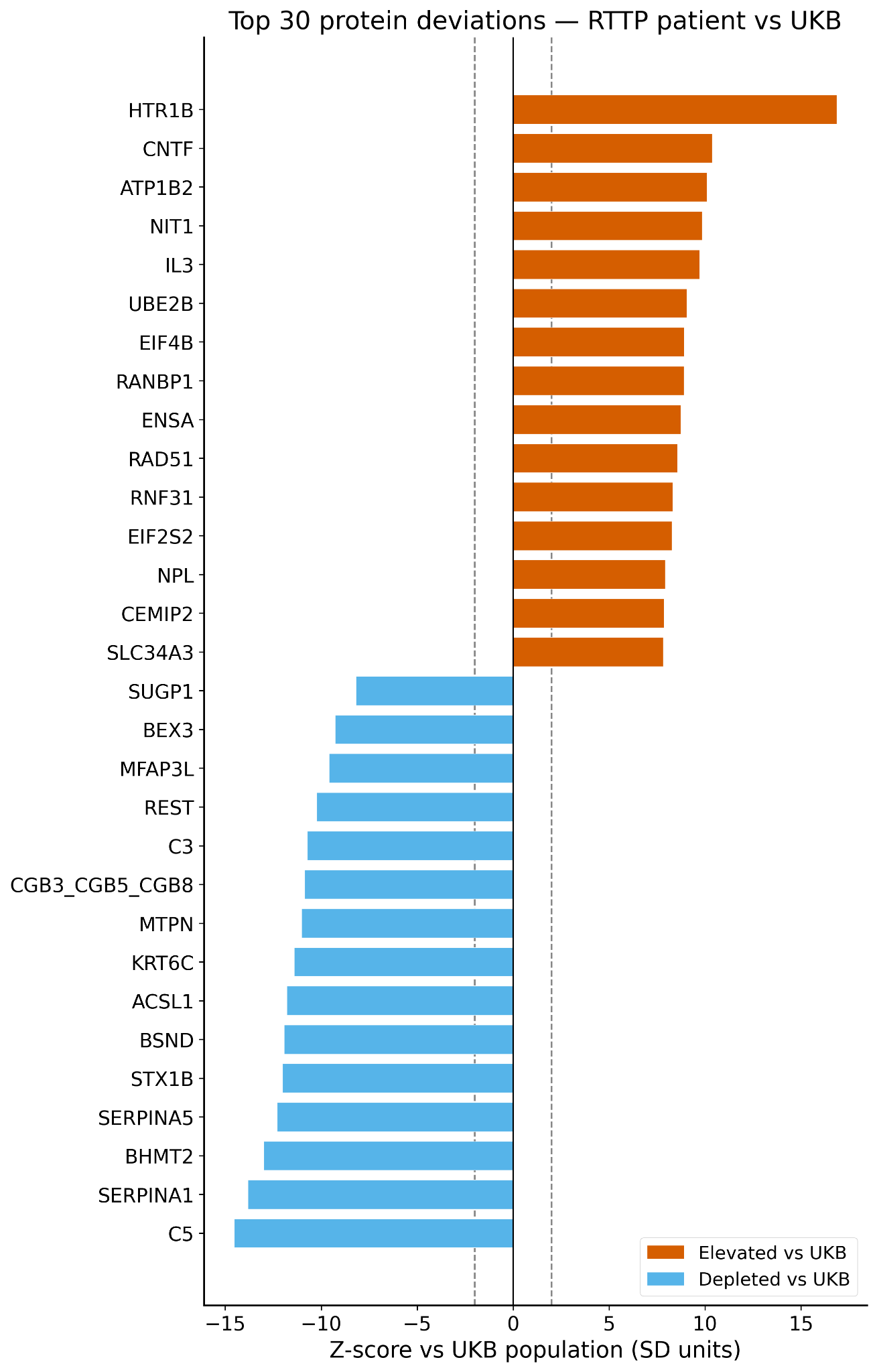
Figure 5. Top protein deviations in the patient relative to the UK Biobank population. Horizontal bars show per-protein Z-scores (SD units); orange, elevated; blue, depleted. Dashed lines mark ±2 SD. Elevated proteins include IL3 and CEMIP2; depleted proteins include complement C3, SERPINA1, and SERPINA5, consistent with immune dysregulation and proteostatic/trafficking stress.
\4. Discussion
4.1 Key Findings
This case documents an exceptionally long-surviving patient with biallelic ITGA3 variants and ILNEB syndrome who has reached his early thirties, far beyond the typical infantile course. The dominant manifestation is a mixed pulmonary phenotype combining conducting-airway disease (bronchiolitis obliterans–like histology) and progressive alveolar fibrosis, accompanied by renal disease requiring transplantation and comparatively mild cutaneous involvement. A prominent fibroinflammatory component—lymphoid aggregates, M2 macrophage enrichment, and reduced regulatory T cells—was evident in tissue and clinically mirrored by stepwise deterioration after respiratory infections. Genomic analysis identified a complex variant landscape at the ITGA3 locus, including nine phased SNVs and a ~1.6 kb intragenic insertion, with marked transcript isoform disruption.
4.2 Mechanistic Insights
The molecular findings support a coherent chain linking genotype to phenotype. The ~1.6 kb intragenic insertion is a strong candidate loss-of-function allele that, alongside the phased coding variants, is expected to compromise normal ITGA3 processing. Because the integrin α3 subunit must be glycosylated, dimerized with β1, and proteolytically matured as it transits the ER and Golgi, defective alleles plausibly produce an ER-to-Golgi trafficking deficit and abnormal protein processing—consistent with the predominance of incomplete-splice-match and non-canonical transcripts and with the ER-weighted proteostatic stress signature. Impaired epithelial adhesion and basement-membrane integrity then destabilize the epithelial barrier, and recurrent epithelial injury—particularly after infection—drives chronic inflammatory remodeling and persistent fibroblast activation, culminating in the fibrosis cascade. Orthogonal organelle- and cell-type-resolved proteomics reinforced this model, extending a single-gene trafficking defect to a systems-level fibroinflammatory signature.
4.3 Comparison with the Literature
The great majority of previously reported ITGA3-related ILNEB cases present in the first months of life and follow a severe, often fatal course, with most patients dying of pulmonary or renal failure before age two. A minority with hypomorphic or atypical variants have survived into later childhood, sometimes with attenuated or organ-restricted disease. The present case sits at the far end of this spectrum: rather than early multi-organ failure, the patient experienced a delayed, slowly progressive course extending into the fourth decade, suggesting preserved residual integrin function sufficient to support prolonged survival while still predisposing to chronic, infection-amplified fibrotic remodeling—a natural history not previously characterized at the molecular level in ILNEB syndrome.
4.4 The Role of Inflammation
A central unresolved question is whether inflammation is a primary driver or a secondary response to epithelial injury. In one interpretation, ITGA3 deficiency primarily destabilizes the epithelial barrier, and the observed infiltrate—lymphoid aggregates, M2 macrophages, and reduced regulatory T cells—arises secondarily from repeated injury and impaired repair. In the other, intrinsic immune dysregulation associated with integrin dysfunction and proteostatic stress actively amplifies profibrotic signaling. The patient’s pattern of disproportionate decline and incomplete recovery after respiratory infections implicates viral and other infectious triggers as accelerants. Chronic post-transplant immunosuppression further complicates the picture, since it may have altered immune homeostasis and the response to infection. These possibilities are not mutually exclusive; the available data cannot establish causal direction, and the interpretation remains hypothesis-generating.
4.5 Clinical Implications
Several practical implications follow. First, this case argues for a broader diagnostic index of suspicion for ITGA3-related disease in older patients with otherwise unexplained combined airway and fibrotic lung disease, especially with renal or cutaneous findings. Second, given the stepwise, infection-associated decline, close monitoring and proactive management around respiratory infections may help preserve pulmonary reserve. Third, the mechanism suggests candidate therapeutic directions rather than established treatments: anti-fibrotic agents to slow parenchymal remodeling, immune modulation to interrupt the fibroinflammatory cycle, and, more speculatively, strategies aimed at the underlying protein-trafficking defect. These are hypotheses for future evaluation and not validated interventions; management should remain individualized and multidisciplinary.
4.6 Limitations
This study has important limitations. It describes a single patient, constraining generalizability, and no technical replicates were available for the genomic and transcriptomic analyses. The proteomic and organelle-resolved signals were derived in part from peripheral samples used as a proxy for lung tissue, which may not faithfully represent compartment-specific biology. The proposed mechanism—linking trafficking deficits to inflammation and fibrosis—was not functionally validated, and the candidate ~1.6 kb insertion and non-canonical transcripts were not experimentally confirmed at the protein level. The contribution of chronic immunosuppression cannot be disentangled from intrinsic disease biology. Accordingly, the mechanistic model should be regarded as hypothesis-generating.
4.7 Future Directions
Future work should prioritize functional validation of the identified variants, including confirmation of the candidate loss-of-function insertion and characterization of the non-canonical splice variants and their effects on protein expression, localization, and ER-to-Golgi transport. Patient-derived organoid and induced pluripotent stem cell models would allow direct interrogation of the trafficking deficit and of strategies to restore integrin maturation or to reprogram activated fibroblasts toward a quiescent state. Continued development of multi-omics integration pipelines—combining genomic, transcriptomic, proteomic, organelle-, cell-type-, and tissue-resolved data—offers a route to test, in a unified framework, whether ITGA3 dysfunction drives trafficking deficits, chronic inflammation, fibroblast activation, and accelerated biological aging.
\5. Conclusion
This case extends the recognized phenotypic spectrum of ILNEB syndrome into the fourth decade, documenting a long-surviving patient with biallelic ITGA3 variants whose dominant manifestation is a mixed conducting-airway and alveolar fibrotic lung disease. Three translational takeaways emerge:
Prolonged survival in ILNEB syndrome is possible and is plausibly enabled by residual integrin function, broadening the clinical picture clinicians should consider in unexplained combined airway-and-fibrotic lung disease with renal or cutaneous features.
Integrated genomic, transcriptomic, and proteomic data converge on a candidate mechanism in which compromised integrin processing produces ER-to-Golgi trafficking and proteostatic stress, linking epithelial fragility to chronic inflammation and progressive fibrosis.
As a hypothesis-generating single case, the findings nominate anti-fibrotic therapy, immune modulation, trafficking-directed strategies, and post-infection surveillance as priorities for functional validation in patient-derived models—illustrating the translational value of a multimodal approach to rare disease.
Supplementary Material
The following supplementary figures present the full organelle-, compartment-, cell-type-, and tissue-resolved proteomic atlas underlying the systems-level analysis summarized in Section 3.5. All panels compare the patient against the UK Biobank (UKB) reference population using the multimodal aging framework (mAge).
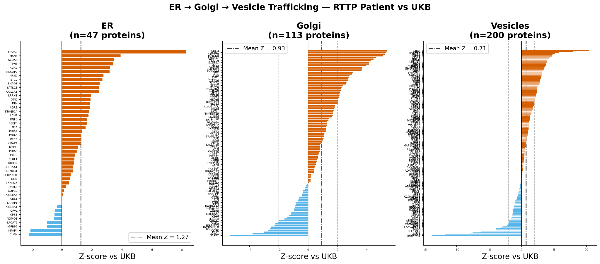
Figure S1. ER→Golgi→vesicle trafficking proteins (patient vs UKB). Per-protein Z-scores across the endoplasmic reticulum (n=47), Golgi (n=113), and vesicle (n=200) compartments; the ER compartment shows the highest mean deviation (mean Z = 1.27), indicating secretory-pathway proteostatic stress.
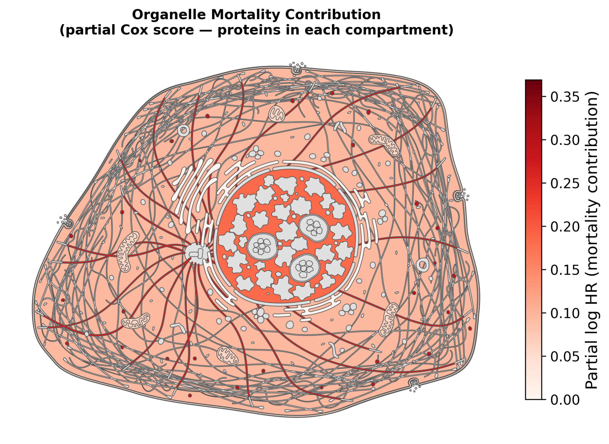
Figure S2. Organelle mortality contribution. Compartment-resolved partial Cox contributions (log-HR) projected onto a schematic cell, highlighting trafficking- and organelle-associated proteins as contributors to mortality-associated proteomic signal.
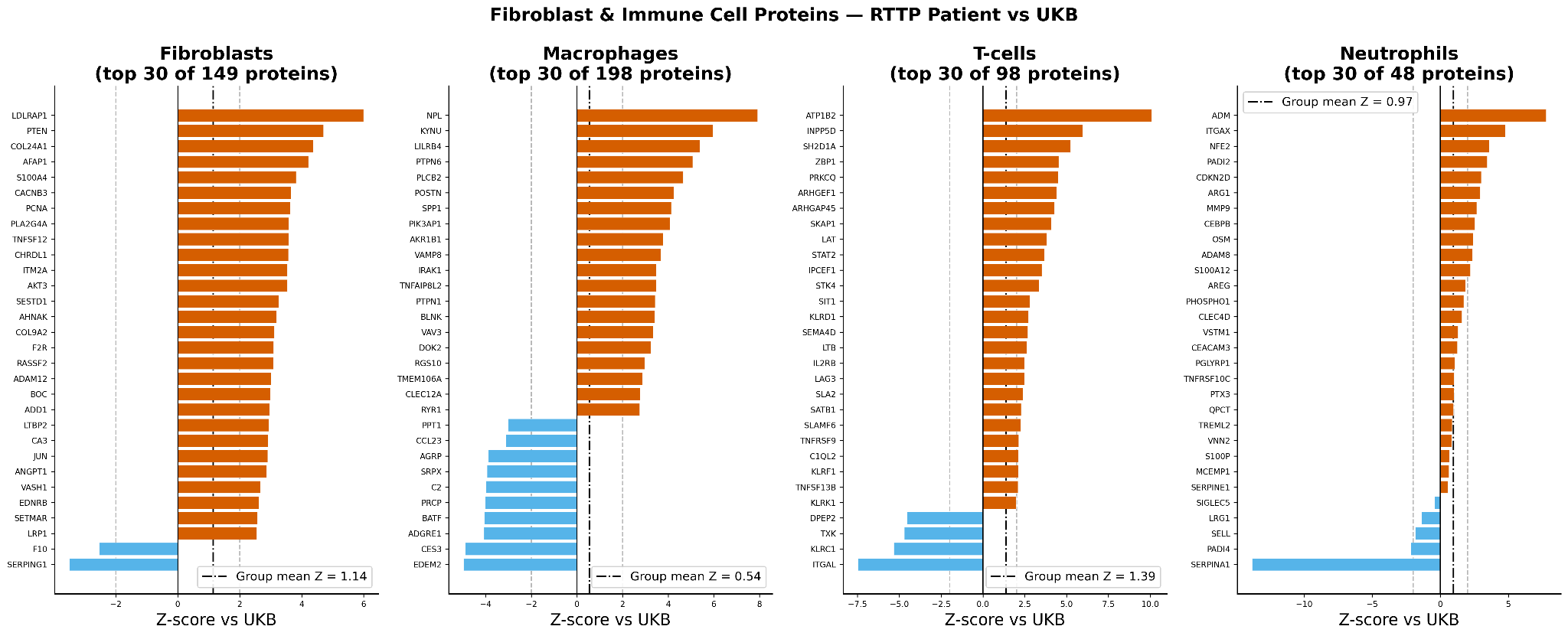
Figure S3. Cell-type-associated protein programs (patient vs UKB). Fibroblast, macrophage, T-cell, and neutrophil protein programs are broadly activated, with the largest shifts in T-cell and fibroblast signatures, supporting inflammation-driven tissue remodeling.
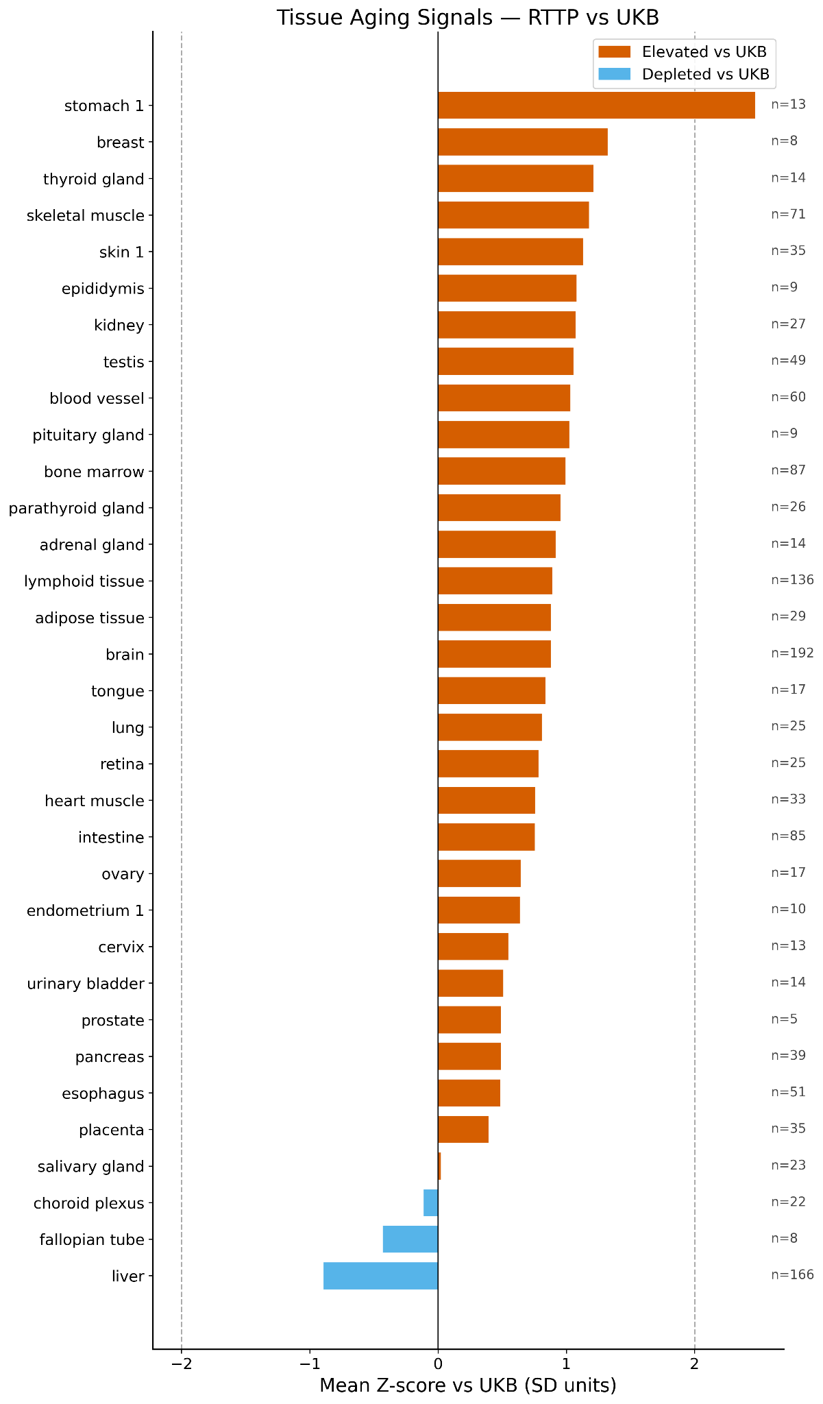
Figure S4. Cell-type aging signals across immune, stromal, endothelial, and epithelial populations, showing widespread multi-cell-type activation consistent with systemic inflammatory and tissue-remodeling stress.
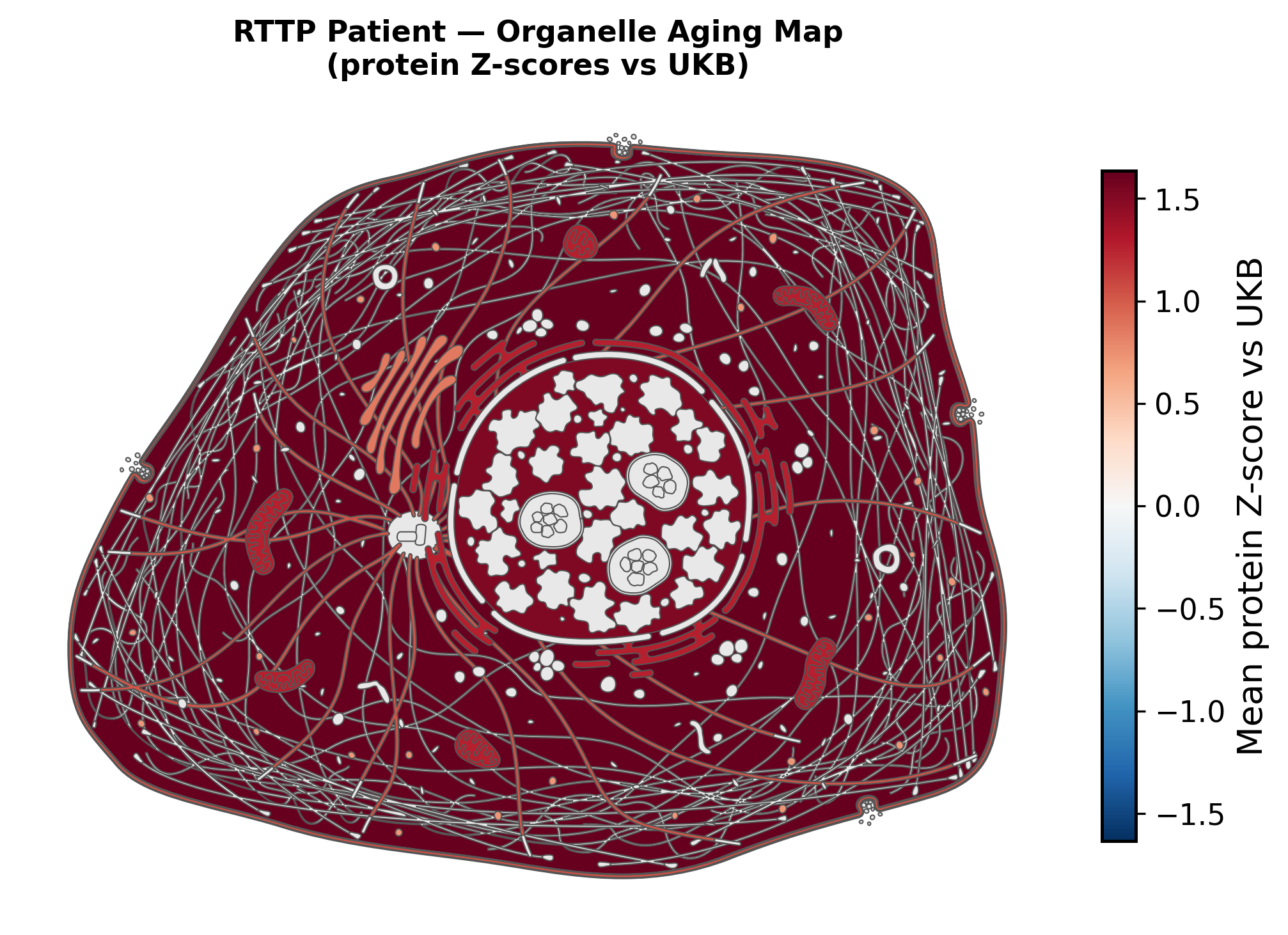
Figure S5. Organelle aging map (protein Z-scores vs UKB). Spatial organelle representation showing broadly elevated aging-associated proteomic signal across intracellular compartments.
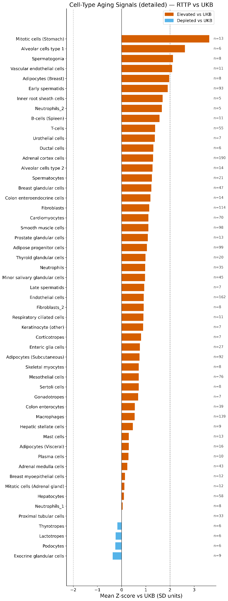
Figure S6. Extended organelle-associated protein deviations (patient vs UKB), ranked by Z-score, complementing the ER/Golgi/vesicle compartment analysis.
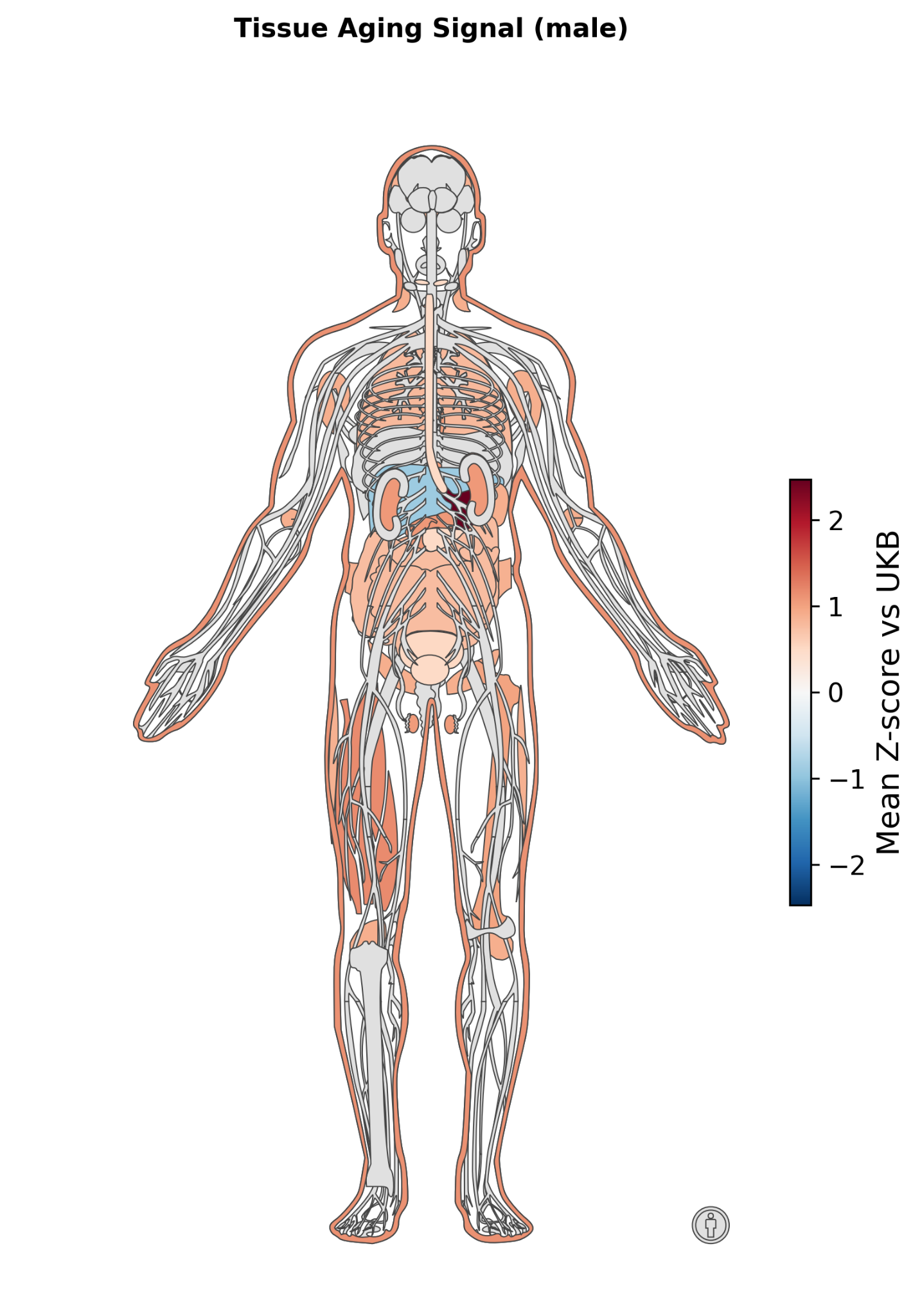
Figure S7. Body-wide tissue aging signal (mean protein Z-score vs UKB), with elevations across gastrointestinal, vascular, muscular, endocrine, and immune-associated tissues.
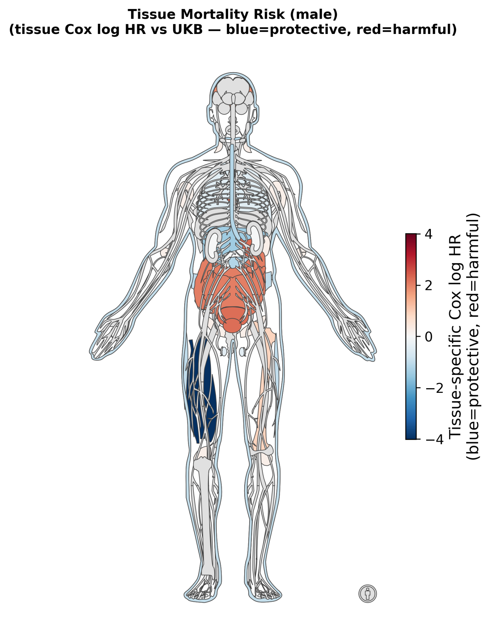
Figure S8. Tissue-specific mortality risk (tissue Cox log-HR vs UKB; blue = protective, red = harmful), showing heterogeneous risk concentrated in abdominal, gastrointestinal, endocrine, and urogenital tissues.
Be the first to comment on this publication.