Purpose
Background: Autoimmune and cardiovascular diseases are diagnostically complex, especially when multiple rare disorders co-occur in a single patient. This study presents a comprehensive genomic analysis of a 50-year-old female patient with mixed ancestry and a history of thrombotic thrombocytopenic purpura (TTP), systemic lupus erythematosus (SLE), and left ventricular non-compaction cardiomyopathy (LVNC).
Methods: Whole genome sequencing (WGS) data was analyzed, and followed by a tiered variant prioritization strategy using the SEQ platform. A symptom-guided filtering approach was applied using Human Phenotype Ontology (HPO) terms associated with each of the patient’s conditions. Additional layers of interpretation included ACMG classification, minor allele frequency thresholds, and pathway enrichment analysis via g:Profiler.
Results: Out of 5.9 million single nucleotide variants (SNVs), 209 of them passed initial filtering based on tier-based classification and population allele frequency (<1%). Furthermore, 42 candidate variants across 25 genes met ACMG criteria (excluding benign categories) and HPO-based phenotype filters aligned with the patient’s symptoms. Several variants were classified as VUS, yet mapped to symptom-relevant genes such as DOCK11, C4A, C4B, ADA2, and DNASE1, indicating potential modifier or overlapping effects between SLE and TTP. Notably, TTN, RPL3L, and GET3 variants aligned with the patient’s cardiac features, while HLA gene variants appeared relevant to her autoimmune and thrombotic manifestations. SV overlaps across SLE, TTP, and LVNC are consistent with convergent mechanisms but lack statistical validation in this single patient case.
Conclusion: This study demonstrates feasibility of a symptom-to-variant filtering strategy to generate phenotype-aligned candidate variants in a diagnostically challenging case. While many of the identified variants are classified as VUS, the integrated analysis of SNVs, SVs, and functional enrichment highlights candidate genes for further investigation. The findings support a polygenic or synergistic model underlying the patient’s complex presentation, requiring follow-up validation and testing across broad cohort studies.
Full paper download: please contact, christine@researchtothepeople.org
Introduction
Autoimmune diseases represent a diagnostic and therapeutic challenge due to their heterogeneous clinical presentations, overlapping symptoms, and multifactorial etiologies involving both genetic predisposition and environmental triggers.1–3 Advances in genomic technologies, particularly next-generation sequencing (NGS), provides a crucial utility for deeper exploration of the genetic contributions to these complex conditions. Although NGS technologies such as whole genome sequencing (WGS) are now more accessible, interpretation of sequencing data remains limited in complex cases, particularly when disease phenotypes are atypical, rare, or incompletely characterized.4–6
This case involves a 50-year-old female patient of mixed Filipino and Caucasian ancestry with overlapping rare autoimmune and cardiovascular conditions. Her clinical history began at age five with an episode of epiglottitis, marking the onset of systemic immune dysregulation. Over the subsequent decades, she was diagnosed with thrombotic thrombocytopenic purpura (TTP), systemic lupus erythematosus (SLE), autoimmune autonomic ganglionopathy (AAG), and left ventricular non-compaction cardiomyopathy (LVNC). TTP was diagnosed at the age of 14 and followed a relapsing course, requiring hundreds of therapeutic plasma exchange treatments with fresh frozen plasma to manage recurrent episodes. The multifaceted nature of her conditions, coupled with persistent autoimmune features, raised the possibility of underlying genetic and immunological contributions. Therefore, WGS was obtained to investigate potential molecular mechanisms driving disease susceptibility, chronicity, and co-occurrence.7
Using WGS data from the patient, a gene-based variant prioritization strategy was previously applied by Shen and colleagues, revealing several rare pathogenic and likely pathogenic variants across multiple genes.7 While that analysis provided foundational insight into possible genetic contributors, it primarily focused on predefined gene-disease associations. In this follow-up study, we expanded upon those findings by applying a symptom-driven variant filtering framework, querying symptoms relevant to SLE, TTP, and LVNC. This approach is based on the premise that clinical symptoms represent the phenotypic outcomes of underlying genetic variations. Therefore, integrating symptom-based filtering can reveal variants that contribute to overlapping or modifying effects across related conditions.8–9
In this approach, we mapped the patient’s symptoms to Human Phenotype Ontology (HPO) terms.10 This is combined with a tiered prioritization strategy as described by Genomize,11 and established classification criteria from the American College of Medical Genetics and Genomics (ACMG).12 Together, these layers enabled us to re-rank variants based not only on predicted pathogenicity but also on clinical relevance to the patient’s presentation, thereby retaining candidate variants potentially missed in prior gene-centric analyses.13
Methods
Patient Sample and Sequencing
Genomic DNA was extracted from a saliva sample obtained from a 50-year-old female patient. WGS was performed using the Illumina platform with PCR-free library preparation. Raw sequencing data were delivered in FASTQ format, as previously described,7 for downstream analysis.
Data Processing and Variant Calling
Sequencing data were processed and analyzed using the SEQ platform (Genomize Bilisim ve Biyoteknoloji AS, Istanbul, Turkey), a cloud-based, AI-supported system designed for clinical-grade NGS analysis.14 The platform integrates public and proprietary databases to enable dynamic, phenotype-driven variant interpretation, including HPO, gnomAD, dbSNP, ACMG and ClinVar.10, 12, 15–17 A complete list of the resources and database versions used during the analysis is available in the Supplementary Methods.
Annotated variants were first categorized using the SEQ platform’s five-tier prioritization framework, which integrates clinical and population-level data to rank variants by predicted pathogenicity and phenotypic relevance.11 For downstream analysis, only Tier I-III variants (high-priority or disease-relevant candidates) were retained. Variants classified as Tier IV or V, which typically represent benign or low-confidence findings, were deprioritized to improve interpretive focus.
Symptom-to-Variant Filtering Strategy
To improve the precision of variant prioritization, we employed a symptom-guided strategy. Symptom-specific virtual gene panels were constructed using relevant HPO terms mapped to the patient’s three core disease domains: (i) SLE, (ii) TTP, and (iii) LVNC. Relevant HPO terms are detailed in Supplementary Methods. Briefly, we employed a lenient keyword-based filtering strategy for HPO terms, and identified those containing the phrases “lupus”, “thrombo”, and “left ventricular”. This initial filtering was intended to capture a broad phenotype spectrum and avoid early exclusion of potentially relevant features in this complex case.
Additional criteria for filtering the variants were based on:
- ACMG classification, including pathogenic (P), likely pathogenic (LP), variants of uncertain significance (VUS), and subcategories such as VUS+ and VUS++.12Particularly, VUS+ and VUS++ criteria represent the variants with some evidence of pathogenicity, but not enough to meet the formal LP criteria.12
- Exonic consequence, focusing on coding variants as defined in Supplementary Methods*.*
- Population frequency thresholds, with a minor allele frequency (MAF) cutoff of <1% across all gnomAD subpopulations.15 This threshold was applied to prioritize rare variants more likely to have functional or clinical impact, while excluding common polymorphisms unlikely to explain a rare or severe phenotype.5, 12, 15, 18
Workflow followed during the analyses for each disease area are detailed in Figure 1.
Pathway and Functional Enrichment Analysis
Functional enrichment analysis was conducted using g:Profiler19 to identify overrepresented Gene Ontology (GO) terms (Biological Process, Molecular Function, and Cellular Component), KEGG and Reactome pathways, and HPO terms.10, 20–21 The input gene list included all prioritized variants for each condition (SLE, TTP, and LVNC) identified in this study. Numbers of input genes and the prioritized variants for these conditions are provided in their respective results sections. The analysis used the g:SCS algorithm to correct for multiple testing, with a significance threshold set at an adjusted p-value < 0.05. Only annotated genes from the Homo sapiens genome were included in the background.19
Results
Overview of Variant Detection
WGS of the patient yielded data suitable for clinical variant interpretation. The sequencing achieved an average depth of 45.51×, with 92.79% of the reference genome covered (5x: 91.91%, 20x: 90.81%, 50x: 43.80%).
Out of 5,900,401 single nucleotide variants (SNVs) initially identified, 209 variants passed tier-based and frequency (<1%) filtering (full list in Supplementary Results). In parallel, 12,127 structural variants (SVs) were detected during initial variant calling.
To further refine candidate variants, we applied a symptom-guided filtering strategy, integrating patient-specific features and HPO terms across three diagnostic categories: SLE, TTP, and LVNC. The analytical workflow is summarized in Figure 1. This follow-up approach extends prior findings and is designed as a feasibility demonstration of symptom-guided filtering in a single complex case.7
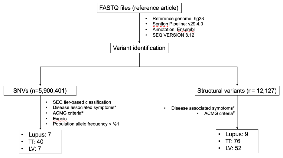
Figure 1. Analysis pipeline and obtained results for SNVs and SVs of the patient’s genome. Starting from the FASTQ file, patient’s WGS data was analyzed by using SEQ platform and promising variants (SNVs and SVs) were identified. *: standardized HPO terms based on three main disease areas (Supplementary Methods), #: Prioritized ACMG classes including VUS, VUS+, VUS++, LP, and P.
Symptom-Guided Filtering: SLE-Related Phenotype
From lupus-related symptom filtering, seven candidate variants were shortlisted (Table 1). Among these, C4A, C4B, and DNASE1 were notable. While classified as VUS under ACMG criteria, they align with known SLE biology and HPO annotations. In addition, immune-deficiency-related genes such as ADA2, CFTR, and DOCK11 were implicated. Interestingly, CFTR, primarily associated with cystic fibrosis, was also annotated with HP:0002725 (systemic lupus erythematosus) in HPO.
| Gene | Consequences | Variant ID | dbSNP | Pathogenicity | Associated Diseases | GT |
|---|---|---|---|---|---|---|
| DOCK11 | Splice Polypyrimidine Tract Variant | chrX:118680470 C->T | rs199582396 | VUS | · XL,XR | Autoinflammatory disease, multisystem, with immune dysregulation · XR | Early-onset immune dysregulation due to dock11 complete deficiency · XR | Early-onset immune dysregulation with autoimmunity due to dock11 partial deficiency | HT |
| C4A | Splice Region Variant | chr6:31991593 C->T | rs879873173 | VUS | · Blood group, rodgers; · AR | C4a deficiency · AD,AR | Autosomal systemic lupus erythematosus · Mu | Behçet disease · Immunodeficiency due to a classical component pathway complement deficiency | HT |
| C4B | Splice Region Variant | chr6:32024331 C->T | rs369924 | VUS | · AR | Complement component 4b deficiency · Immunodeficiency due to a classical component pathway complement deficiency · Systemic lupus erythematosus | HT |
| C1QB | Missense Variant | chr1:22660957 T->C | rs148746097 | VUS | · AR | C1q deficiency · Immunodeficiency due to a classical component pathway complement deficiency | HT |
| CFTR | Missense Variant | chr7:117592032 G->A | rs121908759 | VUS | · AD | Bronchiectasis with or without elevated sweat chloride 1 · AR | Cystic fibrosis · Hypertrypsinemia, neonatal · AD | Pancreatitis, hereditary · Sweat chloride elevation without cf · AR,Mu | Congenital bilateral absence of vas deferens | HT |
| DNASE1 | Missense Variant | chr16:3657022 C->G | rs1799891 | VUS | · AD,AR | Autosomal systemic lupus erythematosus | HT |
| ADA2 | Splice Polypyrimidine Tract Variant | chr22:17191821 AG->A | rs767647109 | VUS | · AR | Vasculitis, autoinflammation, immunodeficiency, and hematologic defects syndrome · AR | Adenosine deaminase 2 deficiency · AD | Diamond-blackfan anemia · AR | Polyarteritis nodosa · AD,AR | Sneddon syndrome | HT |
Table 1. Lupus-Related Variants from Symptom-Guided Analysis
Symptom-Guided Filtering: TTP-Related Phenotype
Symptom-based filtering for thrombotic/thrombocytopenic features yielded 40 variants across 23 genes:
- A homozygous stop-gained/frameshift variant in CBS was classified as LP due to its impact on protein truncation and known association with hyperhomocysteinemia and thrombosis.22–23
- A heterozygous frameshift variant in NKX2-6 (VUS+) has prior links to cardiac malformations and vascular pathology.24–25
Additional variants in TTN, HLA-B, HLA-DQA1, and HLA-DQB1 indicated compound heterozygosity with multiple missense changes, suggesting additive pathogenic potential. Overlap was also noted with six lupus-related genes (DOCK11, C4A, C4B, CFTR, DNASE1, ADA2), hinting at shared mechanisms.
Gene | Consequences | Variant ID | dbSNP | Pathogenicity | Associated Diseases | GT |
CBS | Stop Gained; | chr21:43063074 A->ATGATCTGCAGAGGGCGCGGCTTCAGGGCTCAAGCCCAGCAAAAGCCCCACCTGGATGATCCACCCCAG | - | LP | · AR | Homocystinuria, b6-responsive and nonresponsive types · AR | Thrombosis, hyperhomocysteinemic · AR | Classic homocystinuria · AR | Homocystinuria due to cystathionine beta-synthase deficiency | HM |
NKX2-6 | Frameshift Variant | chr8:23702476 GTGGCTGCCAGA->G | - | VUS+ | · Conotruncal heart malformations · Persistent truncus arteriosus · Common arterial trunk · AR | congenital heart disease · AR | Conotruncal heart malformations · AD,Mu | Tetralogy of fallot | HT |
TCIRG1 | Missense Variant | chr11:68050648 G->A | rs764312517 | VUS | · AR | Osteopetrosis, autosomal recessive 1 · AD | Autosomal dominant severe congenital neutropenia ·AR | Autosomal recessive osteopetrosis ·AR,XR | Dysosteosclerosis · AR | Intermediate osteopetrosis | HT |
KARS1 | Missense Variant | chr16:75631490 C->T | rs149772470 | VUS | · AR | Charcot-marie-tooth disease, recessive intermediate, b · AR | Deafness, congenital, and adult-onset progressive leukoencephalopathy · AR | Leukoencephalopathy, progressive, infantile onset, with or without deafness · AR | Autosomal recessive nonsyndromic hearing loss 89 · AR | Early-onset progressive leukoencephalopathy-central nervous system calcification-deafness-visual impairment syndrome · AR | Rare autosomal recessive non-syndromic sensorineural deafness type dfnb | HT |
DOCK11 | Splice Polypyrimidine Tract Variant | chrX:118680470 C->T | rs199582396 | VUS | · XL,XR | Autoinflammatory disease, multisystem, with immune dysregulation · XR | Early-onset immune dysregulation with autoimmunity due to dock11 partial deficiency | HT |
C4A | Splice Region Variant | chr6:31991593 C->T | rs879873173 | VUS | · Blood group, rodgers · AR | C4a deficiency · AD,AR | Autosomal systemic lupus erythematosus · Mu | Behçet disease · AR | Complement component 4a deficiency · Immunodeficiency due to a classical component pathway complement deficiency · Systemic lupus erythematosus | HT |
C4B | Splice Region Variant | chr6:32024331 C->T | rs369924 | VUS | · C4b deficiency · AR | Complement component 4b deficiency · Immunodeficiency due to a classical component pathway complement deficiency · Systemic lupus erythematosus | HT |
CFTR | Missense Variant | chr7:117592032 G->A | rs121908759 | VUS | · AD | Bronchiectasis with or without elevated sweat chloride 1 · AR | Cystic fibrosis · Hypertrypsinemia, neonatal · AD | Pancreatitis, hereditary · Sweat chloride elevation without cf · AR,Mu | Congenital bilateral absence of vas deferens · AD,AR,XR | Male infertility with azoospermia or oligozoospermia due to single gene mutation | HT |
TTN | Missense Variant | chr2:178552794 C->A | rs959063938 | VUS | · AD | Cardiomyopathy, dilated, 1g · AD | Cardiomyopathy, familial hypertrophic, 9 · AR | Congenital myopathy 5 with cardiomyopathy · AR | Muscular dystrophy, limb-girdle, autosomal recessive 10 · AD | Myopathy, myofibrillar, 9, with early respiratory failure · AD | Tibial muscular dystrophy, tardive · AD | Arrhythmogenic right ventricular cardiomyopathy · AR | Autosomal recessive centronuclear myopathy · Classic multiminicore myopathy · AR | Early-onset myopathy with fatal cardiomyopathy · AD | Familial atrial fibrillation · Familial isolated arrhythmogenic ventricular dysplasia, biventricular form /left dominant form /right dominant form · AD,AR,M,XR | Familial isolated dilated cardiomyopathy · AD | Hereditary myopathy with early respiratory failure · AD | Hypertrophic cardiomyopathy · Inherited isolated arrhythmogenic cardiomyopathy, dominant-left variant /dominant-right variant /biventricular variant · AD | Myopathy, myofibrillar, 9, with early respiratory failure · AR | Titin-related limb-girdle muscular dystrophy r10 · AR | Ttn-related myopathy | c. HT |
TTN | Missense Variant | chr2:178563172 C->T | rs1027584774 | VUS | c. HT | |
TTN | Missense Variant; | chr2:178688679 T->C | rs976519701 | VUS | c. HT | |
TTN | Missense Variant | chr2:178528761 T->C | rs781108499 | VUS | c. HT | |
HLA-B | Splice Polypyrimidine Tract Variant | chr6:31356966 G->C | rs41563412 | VUS | · Mu | Ankylosing spondylitis, susceptibility to, 1 · Severe cutaneous adverse reaction, susceptibility to · Stevens-johnson syndrome, susceptibility to · Toxic epidermal necrolysis, susceptibility to · Mu | Behçet disease · Mu | Giant cell arteritis · Mu | Reactive arthritis · Takayasu arteritis | c. HT |
HLA-B | Frameshift Variant | chr6:31356824 C->CA | rs9281379 | VUS | c. HT | |
HLA-B | Missense Variant | chr6:31356280 C->A | rs12697943 | VUS | c. HT | |
HLA-B | Missense Variant | chr6:31356822 T->G | rs1050538 | VUS | c. HT | |
HLA-DQA1 | Missense Variant | chr6:32641456 T->C | rs3188043 | VUS | · AR,Mu | Celiac disease, susceptibility to · Mu | Adult-onset myasthenia gravis · AR | Idiopathic achalasia | c. HT |
HLA-DQA1 | Missense Variant | chr6:32641444 G->C | rs12722061 | VUS | c. HT | |
HLA-DQA1 | Missense Variant | chr6:32641450 A->C | rs3207985 | VUS | c. HT | |
HLA-DQA1 | Missense Variant | chr6:32641436 G->A | rs3207983 | VUS | c. HT | |
HLA-DQA1 | Missense Variant | chr6:32641445 A->T | rs3208105 | VUS | c. HT | |
HLA-DQA1 | Missense Variant | chr6:32641502 C->T | rs9272709 | VUS | c. HT | |
HLA-DQA1 | Missense Variant | chr6:32641451 G->A | rs28383449 | VUS | c. HT | |
HLA-DQA1 | Missense Variant | chr6:32641454 A->G | rs12722069 | VUS | c. HT | |
HLA-DQA1 | Missense Variant | chr6:32641501 G->C | rs1142335 | VUS | c. HT | |
HLA-DQB1 | Missense Variant | chr6:32664926 G->A | rs9274395 | VUS | · AR,Mu | Celiac disease, susceptibility to · AD | Creutzfeldt-jakob disease, variant, resistance to · Mu | Multiple sclerosis, susceptibility to · AR | Idiopathic achalasia · Narcolepsy type 1 · Narcolepsy type 2 | c. HT |
HLA-DQB1 | Missense Variant | chr6:32664992 T->C | rs767838657 | VUS | c. HT | |
HLA-DQB1 | Missense Variant | chr6:32664971 T->A | rs750855857 | VUS | c. HT | |
HLA-DQB1 | Missense Variant | chr6:32664993 A->T | rs281862065 | VUS | c. HT | |
DNASE1 | Missense Variant | chr16:3657022 C->G | rs1799891 | VUS | · AD | Systemic lupus erythematosus | HT |
SCN11A | Splice Polypyrimidine Tract Variant | chr3:38946916 C->CA | rs552650164 | VUS | · AD | Episodic pain syndrome, familial, 3 · AD | Hereditary sensory and autonomic neuropathy · AD | Familial episodic pain syndrome with predominantly lower limb involvement · AD | Hereditary sodium channelopathy-related small fibers neuropathy | HT |
SEC61A1 | Splice Polypyrimidine Tract Variant | chr3:128066940 G->A | rs773757716 | VUS | · AD | Immunodeficiency, common variable, 15 · AD | Neutropenia, severe congenital, 11 · AD | Tubulointerstitial kidney disease, autosomal dominant, 5 · AD | Hyperuricemic nephropathy, familial juvenile type 4 · AD | Sec61a1 deficiency | HT |
TBXAS1 | Missense Variant | chr7:139953397 C->A | rs5768 | VUS | · AR | Ghosal hematodiaphyseal syndrome | HT |
CUBN | Splice Polypyrimidine Tract Variant | chr10:16835211 G->T | rs190546478 | VUS | · AR | Imerslund-grasbeck syndrome · AR | Proteinuria, chronic benign | HT |
RPL3L | Missense Variant | chr16:1946720 G->A | rs374470658 | VUS | · AR | Cardiomyopathy, dilated, 2d · AR | Dilated cardiomyopathy · AD,AR,M,XR | Familial isolated dilated cardiomyopathy | HT |
DTNBP1 | Splice Region Variant; | chr6:15593088 G->GAA | rs199770715 | VUS | · AR | Hermansky-pudlak syndrome 7 · Hermansky-pudlak syndrome due to bloc-1 deficiency | HT |
ADA2 | Splice Polypyrimidine Tract Variant | chr22:17191821 AG->A | rs767647109 | VUS | · AD,AR | Sneddon syndrome · AR | Vasculitis, autoinflammation, immunodeficiency, and hematologic defects syndrome · AR | Adenosine deaminase 2 deficiency · AD | Diamond-blackfan anemia · AR | Polyarteritis nodosa | HT |
GET3 | Missense Variant | chr19:12737533 G->T | rs749041448 | VUS | · AR | Cardiomyopathy, dilated, 2h · AD,AR,M,XR | Familial isolated dilated cardiomyopathy | HT |
GBA1 | Missense Variant; | chr1:155236246 G->A | rs75548401 | VUS | · AR | Gaucher disease, perinatal lethal · AD | Lewy body dementia, susceptibility to · AD,Mu | Parkinson disease, late-onset, susceptibility to · Fetal gaucher disease · AR | Gaucher disease · AR | Gaucher disease type 1 · AR | Gaucher disease type 2 · AR | Gaucher disease type 3 · AR | Gaucher disease-ophthalmoplegia-cardiovascular calcification syndrome · AD | Parkinson disease | HT |
GALC | Missense Variant | chr14:87986597 T->C | rs147313927 | VUS | · AR | Krabbe disease · Adult krabbe disease · Infantile krabbe disease · Late-infantile/juvenile krabbe disease | HT |
Table 2. Variants associated with TTP-related phenotypes.
Symptom-Guided Filtering: LVNC-Related Phenotype
Seven variants were associated with LVNC-related HPO terms. Of these:
- Four variants resided in TTN, a key gene encoding titin and heavily implicated in cardiomyopathies and arrhythmias.26
- RPL3L and GET3 also emerged with cardiomyopathy annotations.27–28
- All variants caused missense substitutions and were classified as VUS, highlighting uncertain but plausible relevance.
Notably, six of the seven LVNC-related variants were shared with the TTP gene set, potentially reflecting on a phenotypic and genetic overlap between the two conditions.
Gene | Consequences | Variant ID | dbSNP | Pathogenicity | Associated Diseases | GT |
TTN | Missense Variant | chr2:178552794 C->A | rs959063938 | VUS | · AD | Cardiomyopathy, dilated, 1g · AD | Cardiomyopathy, familial hypertrophic, 9 · AR | Congenital myopathy 5 with cardiomyopathy · AR | Muscular dystrophy, limb-girdle, autosomal recessive 10 · AD | Myopathy, myofibrillar, 9, with early respiratory failure · AD | Tibial muscular dystrophy, tardive · AD | Arrhythmogenic right ventricular cardiomyopathy · AR | Autosomal recessive centronuclear myopathy · Classic multiminicore myopathy · AR | Early-onset myopathy with fatal cardiomyopathy · AD | Familial atrial fibrillation · Familial isolated arrhythmogenic ventricular dysplasia, biventricular form /left dominant form /right dominant form · AD,AR,M,XR | Familial isolated dilated cardiomyopathy · AD | Hereditary myopathy with early respiratory failure · AD | Hypertrophic cardiomyopathy · Inherited isolated arrhythmogenic cardiomyopathy, dominant-left variant /dominant-right variant /biventricular variant · AD | Myopathy, myofibrillar, 9, with early respiratory failure · AR | Titin-related limb-girdle muscular dystrophy r10 · AR | Ttn-related myopathy | c.HT |
TTN | Missense Variant | chr2:178563172 C->T | rs1027584774 | VUS | c.HT | |
TTN | Missense Variant; | chr2:178688679 T->C | rs976519701 | VUS | c.HT | |
TTN | Missense Variant | chr2:178528761 T->C | rs781108499 | VUS | c.HT | |
RPL3L | Missense Variant | chr16:1946720 G->A | rs374470658 | VUS | · AR | Cardiomyopathy, dilated, 2d · AR | Dilated cardiomyopathy · AD,AR,M,XR | Familial isolated dilated cardiomyopathy | HT |
GET3 | Missense Variant | chr19:12737533 G->T | rs749041448 | VUS | · AR | Cardiomyopathy, dilated, 2h · AD,AR,M,XR | Familial isolated dilated cardiomyopathy | HT |
AIP | Missense Variant | chr11:67483205 G->A | rs145047094 | VUS | · AD,SMu | Pituitary adenoma 1, multiple types · Acromegaly · AD | Growth hormone secreting pituitary adenoma 1 · AD,XD | Pituitary gigantism · AD | Prolactinoma · Silent pituitary adenoma | HT |
Table 3. LVNC-related variants identified via HPO symptom filtering.
Structural Variants (SV)
SV analysis identified:
- 9 SLE-related, 76 TTP-related, and 52 LVNC-related SVs (Figure 2; Supplementary Results).
- We observed 7 and 17 SV overlaps respectively; these counts are descriptive and consistent with shared mechanisms but not statistically tested.
- One lupus-specific duplication (IGHG2, MIR8071-2, COPDA1) implicates regulatory disruption of humoral immune response and immune signaling.
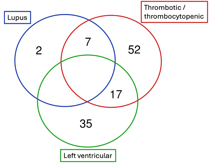
Figure 2. Venn Diagram showing distinct and mutual SVs across patient’s phenotypes. SVs among SLE, TTP, and LVNC phenotypes are represented with blue, red and green circles, respectively. No overlapping SVs between SLE and LVNC features are observed.
| Position | SV Type | Gene/Transcript Info | Pathogenicity |
|---|---|---|---|
| chr14 105630632-105649462*** | DUP | IGHG2, ENST00000641095, Exon 1-6 MIR8071-2, ENST00000616772, Exon 1 COPDA1, ENST00000497397, Exon 1-3 | VUS |
| chr16 88646281-88646336*** | DUP | CYBA, ENST00000261623, Intron 4 | VUS |
| chr22 17184893-17184948 | DEL | ADA2, ENST00000399837, Intron 7 | VUS |
| chrX 118494897-118494967 | DEL | DOCK11, ENST00000276202, Upstream (-848) | VUS |
| chr2 203873328-203873369 | DEL | CTLA4, ENST00000648405, Exon 4 | VUS |
| chr4 101556664-101556697 | DEL | BANK1, ENST00000504592, Intron 2 | VUS |
| chr11 128478355-128478381 | DEL | ETS1, ENST00000392668, Intron 8 | VUS |
| chr13 102652398-102652435 | DEL | TPP2, ENST00000376052, Intron 24 | VUS |
| chr19 33458552-33458580 | DEL | PEPD, ENST00000244137, Intron 9 | VUS |
Table 4. Lupus-Thrombotic/Thrombocytopenic
*: Variants that are unique for lupus-related features only
| Position | SV Type | Gene/Transcript Info | Pathogenicity |
|---|---|---|---|
| chr1 236755585-236755675 | DEL | ACTN2, ENST00000366578, Intron 17 | VUS |
| chr12 21863283-21863328 | DEL | ABCC9, ENST00000261200, Intron 19 ENSG00000257022, ENST00000539874, Intron 2 | VUS |
| chr15 71199892-71199918 | DEL | THSD4, ENST00000261862, Intron 3 | VUS |
| chr5 156187513-156187555 | DEL | SGCD, ENST00000517913, Intron 3 | VUS |
| chr11 2458634-2458661 | DEL | KCNQ1, ENST00000155840, Intron 1 | VUS |
| chr11 2699488-2699514 | DEL | KCNQ1, ENST00000155840, Intron 11 KCNQ1OT1, ENST00000597346, Exon 1 | VUS |
| chr10 68193811-68193840 | DEL | MYPN, ENST00000358913, Intron 13 | VUS |
| chr4 47617955-47617984 | DEL | CORIN, ENST00000273857, Intron 19 | VUS |
| chr2 43835238-43835264 | DEL | ABCG5, ENST00000405322, Intron 2 | VUS |
| chr11 19197365-19197396 | DEL | CSRP3, ENST00000265968, Intron 1 CSRP3-AS1, ENST00000527978, Intron 1 | VUS |
| chrX 32554865-32554900 | DEL | DMD, ENST00000357033, Intron 16 | VUS |
| chrX 32601661-32601690 | DEL | DMD, ENST00000357033, Intron 12 | VUS |
| chr19 38539893-38539920 | DEL | RYR1, ENST00000359596, Intron 84 | VUS |
| chr20 44162766-44162791 | DEL | JPH2, ENST00000372980, Intron 1 | VUS |
| chr1 3105928-3181110 | DEL, DUP | PRDM16, ENST00000270722, Intron 1 | VUS |
| chr15 48547722-48547755 | DEL | FBN1, ENST00000316623, Intron 6 | VUS |
| chr12 112442831-112442858 | DEL | PTPN11, ENST00000351677, Intron 1 | VUS |
Table 5. Thrombotic/Thrombocytopenic - Left Ventricular
Functional Enrichment Analysis
To contextualize the biological roles of the prioritized variant-associated genes, we conducted functional enrichment analysis using g:Profiler across multiple biological and phenotypic ontologies including GO, HPO, KEGG, Reactome, and WikiPathways databases.
Analysis of SNV-Associated Genes
We first analyzed the SNV-containing gene set derived from the patient's genome data. This included 24 genes (C4A, C4B, C1QB, HLA-DQB1, DNASE1, HLA-B, ADA2, SEC61A1, GBA1, DOCK11, TCIRG1, CFTR, CBS, CUBN, AIP, SCN11A, TBXAS1, KARS1, GALC, NKX2-6, TTN, RPL3L, GET3, DTNBP1), excluding HLA-DQA1 which was not recognized by g:Profiler.
HPO: A total of 65 significant HPO terms were enriched (Figure 3A). Among the most significant were:
- Abnormality of blood and blood-forming tissues (HP:0001871), all 24 genes matched.
- Abnormality of the cardiovascular system (HP:0001626), 23 genes matched.
- Other HPO terms included oral ulcers, complement system defects, thrombocyte and platelet abnormalities, lupus, and autoantibody positivity.
Upon counting which genes appeared the most across the identified terms, we saw: C4A (annotated to 63 HPO terms), followed by DNASE1 (51), C4B (50), HLA-B (49), and ADA2 (46). Interestingly, CBS and NKX2-6, which were previously flagged as TTP-associated variants, were involved in 21 and 13 HPO terms respectively, related to vasculature and inflammatory features in skin and joints. In addition, cardiovascular-related terms included AIP (18 terms), TTN and RPL3L (11 each), and GET3 (9).
In total, 5 KEGG Pathways terms were enriched, including systemic lupus erythematosus, phagosome, pertussis, Staphylococcus aureus infection and Vibrio cholera infection (Figure 3B). Notably, C4A, C4B, C1QB, and HLA-DQB1 were commonly implicated in immune-related pathways. TCIRG1 and SEC61A1 were enriched in the phagosome-related pathways, suggesting an immunoregulatory axis independent of the complement genes.
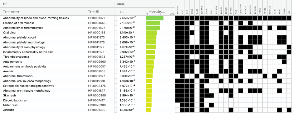

Figure 3. Functional enrichment analyses depicting the top list of (A) HPO terms, and (B) KEGG pathways associate with the genes harboring variants observed in the patients WGS data.
Incorporating SVs, Summary and Comparative Insights
We also conducted a parallel enrichment analysis using genes affected by SVs, which is provided in Supplementary Results. While our main results emphasized SNVs due to their higher resolution and immediate clinical interpretability, the SV-inclusive analysis offered an additional layer to validate and broaden the findings from the SNV-based results. This combined view allowed us to identify shared and distinct patterns in genes, pathways, and ontologies impacted by either variant type, and presented potential contributors that may have been overlooked when analyzing SNVs or SVs in isolation.
The SV gene sets showed enrichment of similar categories including immune dysfunction, platelet and thrombocyte defects, abnormal heart morphology, and autoimmune diseases, consistent with SNV-only analyses - and interpreted as exploratory rather than confirmatory. Several SV-associated genes were enriched for abnormalities in thrombocyte physiology and cardiac structure, in line with the patient’s clinical diagnoses of TTP and LVNC. However, immune-related enrichments, especially autoimmunity and HLA-related pathways, were more prominent in SNV-driven results, likely due to the presence of C4A, C4B, HLA-DQB1, and HLA-B in the SNV set. Looking into both SV- and SNV-based enrichment, we did not observe any additional pathways or ontologies of significance.
Thus, we primarily focused our reporting and discussion on the SNV-based results to maintain clinical interpretability, but we acknowledge that SVs contribute meaningfully to the broader genomic profile. The enrichment patterns remained convergent across both variant types, and are consistent with known immunologic and cardiovascular biology.
Discussion
This study demonstrates the utility of a symptom-to-variant approach for whole genome interpretation in a diagnostically complex case involving multiple rare and autoimmune conditions. Previously, Shen and colleagues performed genomic analysis of this patient, and employed a predefined gene-disease association strategy. The approach identified numerous variants (allele frequency <5%) associated with SLE, TTP, and LVNC when considered as separate disease entities.7 Our current study aimed to complement those findings by shedding light on possible interconnections between diseases and underlying symptoms, rather than treating each in isolation. While doing so, a stringent MAF threshold (<1%) was employed to exclude common polymorphisms that remain limited in explaining rare or severe phenotypes.
In this patient’s case, the majority of variants identified through WGS were classified as VUS, making it challenging to interpret. This is partially also because pathogenicity alone is not sufficient for clinical relevance, particularly in patients with atypical or overlapping phenotypes. For example, genetic pathogenicity may indicate a predisposition to certain pathologies but does not guarantee disease manifestation, as clinical expression can depend on environmental influences or the presence of additional genetic modifiers that may be protective.29–30 Conversely, disease may still develop even when major known genetic drivers appear within normal population ranges.31
By applying ACMG guidelines in combination with symptom-driven filtering, we were able to systematically evaluate and reprioritize variants including VUS according to their potential clinical relevance.12 This framework allowed us to highlight candidate variants that align with the patient’s disease relevant features, including potential modifiers and cross-condition variants that may have been overlooked in strictly disease-focused workflows. However, leveraging VUS also carries the risk of false positives, as many of these variants lack sufficient functional data or clear genotype-phenotype correlations. In particular, the impact of a VUS on gene function, expression, or genomic stability often remains poorly characterized. To mitigate this risk, we applied stringent filtering criteria, prioritizing rare variants based on their genomic location and allele frequency, including variants from incidental findings, especially in the case of single nucleotide variants (SNVs).32–33 Structural variants (SVs), while considered, were filtered with slightly less stringency, as discussed later.
In line with this, heterozygous variants in autosomal recessive (AR) genes were also considered due to their potential clinical relevance. While AR conditions typically require biallelic pathogenic variants for full disease manifestation, numerous studies have reported cases where heterozygous carriers exhibit partial phenotypes or an increased risk of related conditions due to modifier effects, incomplete penetrance, or oligogenic inheritance.4, 34–35 For example, although ADA2, DNASE1, and CTLA4 are generally associated with recessive inheritance, heterozygous variants have been implicated in broader or atypical immune phenotypes.36–37 Moreover, in immune-mediated and cardiomyopathic conditions, heterozygous variants in genes typically associated with autosomal recessive inheritance have been reported to contribute to atypical or subclinical phenotypes, such as in genes ADA2, CTLA4, PTPN11, DSG2, and PKP2.36, 38–41
An additional consideration was given for deep intronic variants and SVs, in support of the SNV based approach, and to account for compound heterozygosity. Accordingly, compound heterozygosity indicates the presence of two distinct pathogenic variants on separate alleles of the same gene. Compound heterozygosity can have causal roles, but standard short-read WGS may detect only one allele due to technical limitations, such as structural variation or coverage gaps.17, 42 Thus, even a single heterozygous variant in a symptom-relevant gene may warrant clinical consideration. In line with this, heterozygous variants and SVs were retained in our analysis when found in genes with known relevance to the patient’s symptoms, especially when rare and classified as P or LP, pathogenic and likely pathogenic, respectively. Moreover, the filtering approach for SNVs was initially confined to exonic variants. This decision was made because the sample size was limited, and drawing assumptions from non-coding regions would have been speculative and difficult to interpret. Therefore, in this context, WGS was not necessarily superior to WES for small variant analysis. However, a primary advantage of using WGS data was the ability to evaluate SVs.
Out of 5.9 million SNVs, we identified 209 variants, despite most being annotated as VUS. We further refined this list using disease-relevant ontologies and ACMG pathogenicity criteria. This allowed a degree of stringency, and presented a list to 42 candidate variants. Two main observations emerged based on these variants: (i) they were distributed across 25 genes which are known to associate with the patient’s disease phenotypes, and (ii) they were shared across the TTP, SLE and LVNC-features. This tempted us to consider whether some of these variants have unknown pathogenicity, and more importantly whether there are shared mechanisms or modifier effects across the patient’s diseases. Most prioritized variants are VUS and are not clinically actionable without orthogonal confirmation (e.g., Sanger sequencing, polymerase chain reaction (PCR), segregation, and functional assays). Therefore, preclinical validations and analyses across large cohort studies are warranted, given that rare and complex nature of her conditions.43 Some of the genes that emerged from the analyses, thus require further attention. For instance, the genes DOCK11, C4A, C4B, CFTR, DNASE1, and ADA2 represented these potential overlapping features between SLE and TTP.36, 44–49
Functional review of these genes supports their roles in immune pathways. DOCK11 encodes a guanine nucleotide exchange factor involved in immune cell signaling, and both SNV and SV findings in this locus may contribute to dysregulated B- and T-cell function observed in lupus.44–45 Complement-related genes including C4A, C4B, C1QB, and C1QC are well-recognized lupus susceptibility loci, with deficiencies in classical complement components strongly linked to impaired immune complex clearance and increased risk of autoimmunity.50 ADA2 deficiency has been associated with systemic vasculopathy and immune dysregulation, and both small variants and intronic SVs in this gene may contribute to the vascular and inflammatory dysfunctions in this patient.36 DNASE1 encodes an endonuclease critical for clearing extracellular DNA; reduced function is a known lupus risk factor due to impaired clearance of apoptotic material and subsequent autoantibody generation.51–52 CFTR, while primarily associated with cystic fibrosis, has been implicated in immune regulation, and variants in this gene may contribute to inflammatory processes that overlap with lupus phenotypes.46–47 Among SVs, common intronic changes were observed in BANK1, ETS1, ADA2, PEPD, and CTLA4, all of which have established or putative roles in B-cell signaling, T-cell regulation, and immune tolerance in lupus pathogenesis.36, 53–59 Of particular note, a lupus-specific duplication was identified spanning IGHG2, MIR8071-2, and COPDA1. Alterations in immunoglobulin heavy chain genes such as IGHG2 may affect humoral responses, while the presence of a microRNA gene (MIR8071-2) and long noncoding RNA (COPDA1) suggests additional layers of post-transcriptional and regulatory effects that could influence immune function.60–63 Hence, the findings suggest convergent mechanisms involving complement pathways and immune cell regulation that may underlie both lupus and thrombotic phenotypes that require functional assays and further data to confirm relevance of the VUS in these cases.43
Several genes identified in this analysis were also found relevant to the patient’s cardiomyopathic and thrombotic phenotypes. TTN, which encodes titin, is an established gene in LVNC and dilated cardiomyopathy, and the presence of multiple variants including compound heterozygous changes suggests that titin dysfunction may contribute significantly to the patient’s cardiac manifestations.26 Supporting this, functional enrichment linked TTN to cardiovascular system abnormalities alongside RPL3L and GET3, both of which are involved in muscle contraction and protein homeostasis, respectively, and have been implicated in cardiomyopathic processes.27–28, 64 In the immune and thrombotic domain, HLA-B, HLA-DQA1, and HLA-DQB1 were strongly represented in the enrichment analysis and are known to influence autoimmune susceptibility and thrombocytopenia, highlighting their potential relevance to the patient’s history of TTP.59, 65–66 Two genes emerged as particularly attractive TTP-associated candidates: CBS, where the patient carries a homozygous stop-gained/frameshift variant classified as LP (likely pathogenic), and NKX2-6, which harbors a heterozygous frameshift variant classified as VUS+. Both were recurrently annotated in enrichment terms related to vascular and inflammatory abnormalities.22–25 In addition, common structural variants were identified in cardiomyopathy-associated genes that code for ion channels, as well as musculoskeletal and connective tissue proteins. These genes include ACTN2, ABCC9, THSD4, SGCD, KCNQ1, MYPN, CORIN, CSRP3, DMD, RYR1, JPH2, PRDM16, and FBN1, though their individual contribution in this case remains uncertain. Together, these findings emphasize that both small variants and compound heterozygous events, particularly in TTN and CBS, along with immune-modulatory loci such as HLA genes, may underlie the overlapping cardiac and thrombotic features observed in this patient. Yet, read-level quality control metrics and loss-of-function confidence scoring (e.g., transcript-level nonsense-mediated decay67) were not provided for the respective variants; confirmations on these metrics and zygosity are pending.
In addition to SLE, TTP, and LVNC, the patient also carries a diagnosis of autoimmune autonomic ganglionopathy (AAG), a rare condition characterized by dysautonomia and neuropathic features. Using phenotype terms related to AAG such as “neuropathy” and “dysautonomia” in functional queries, several genes from the patient’s variant set emerged with potential relevance.68 SCN11A, a sodium channel gene implicated in autonomic dysfunction and sensory neuropathies, represents a plausible candidate for the autonomic features observed.69–70 SEC61A1, encoding a key component of the endoplasmic reticulum translocation machinery, has been linked to immune-related neuropathies and impaired cellular stress responses.71–72 Although no autoimmune role has been observed, lysosomal storage-related genes GBA1 and GALC also appeared in the analysis, consistent with their known associations with neurodegeneration and peripheral nerve dysfunction as in Parkinson’s disease.73–75 Additional hits included KARS1 which has been implicated in neuromuscular disorders,76 as well as immune regulatory genes such as ADA2, HLA-DQB1, HLA-DQA1, and HLA-B, which reinforce the role of autoimmunity in AAG pathogenesis. While these associations remain exploratory, it is tempting to consider these variants may influence neuronal and immune regulatory pathways to contribute to the patient’s autonomic neuropathy, complementing the autoimmune basis of her broader disease spectrum.
A central critique emerging from this case is whether the patient’s presentation reflects a multifactorial interplay of genetic and environmental influences.1, 77 Accordingly, we observed hints at disrupted complement activation, coagulation cascades, and platelets and immune cell functions, which can explain some mechanistic overlaps between SLE, TTP, LVNC, and even aspects of autonomic neuropathy.78–79
Together, these patterns suggest a likely polygenic or synergistic disease model,80 in which variants across interconnected immune, vascular, and cardiac pathways could be additive and may collectively contribute to the complex phenotype observed in this patient.
Limitations and Future Directions
This study builds upon earlier analyses demonstrating the feasibility of symptom-guided variant filtering in a single, diagnostically complex case. Consequently, formal benchmarking against alternative analytical pipelines and validation across larger cohorts were beyond the scope of the current work and will be addressed in future studies.
This study is limited by reliance on existing public variant annotations (e.g., ClinVar, gnomAD), which may underrepresent rare or population-specific alleles. Additionally, most VUS variants, as well as the Tier IIB and V variants in this study, remain difficult to interpret and draw strong associations without broader cohort-level evidence. Sequencing errors and false positive variant calls should be taken into account, particularly for rare or low-frequency variants which can hinder interpretation.
A key finding of this study was a homozygous stop-gained/frameshift variant in CBS, classified as likely pathogenic and central to the thrombotic interpretation. However, this variant, as well as several other prioritized findings, have not yet undergone locus-specific technical validation. In particular, read-level quality control metrics, multi-caller concordance, and high-confidence loss-of-function annotation were not independently verified. Therefore, the robustness of zygosity, variant calling accuracy, and predicted loss-of-function impact require further confirmation before definitive clinical conclusions can be drawn. At the same time, the symptom-based filtering approach employed here has some intrinsic limitations. Mapping clinical HPO terms allowed for a structured, phenotype-driven workflow, but the granularity and accuracy of HPO annotations are variable. Many features overlap across diseases, and broad terms can dilute specificity, leading to challenges in distinguishing disease-specific signals.81–82 While HPO remains a valuable starting point, future analyses may benefit from incorporating clinical coding systems such as ICD-10 or algorithmically defined phenotypes from resources like PheKB.83 These frameworks provide more fine-grained, population-anchored features that could sharpen symptom-driven variant interpretation and reduce ambiguity.
Given the single-case scope of this study, the analyses remain descriptive and hypothesis-generating and necessitate formal statistical evaluation. For example, overlaps of SV across symptom-guided panels require confirmation to determine whether the observed overlap exceeds random expectation given the overall SV burden and panel sizes. Similarly, enrichment results may be influenced by pleiotropic loci (e.g., complement and HLA genes), annotation density, and the choice of background set.84–85 Thus, enrichment should be interpreted as supportive rather than confirmatory, and matched-background or permutation-based testing is warranted. Methodological and analytical future directions aimed at improving the overall workflow include:
• Validating the prioritized variants through orthogonal assays, including Sanger sequencing and PCR.33
• Performing locus-specific validation of high-priority variants (e.g., CBS) by recomputing read-level quality control metrics (depth, allele balance, mapping quality, strand bias), assessing multi-caller concordance, re-annotating across canonical transcripts using Ensembl VEP,86 applying structured ACMG PVS1 criteria,67 and conducting ancestry-aware population frequency checks to strengthen loss-of-function evidence and confirm zygosity.15, 87–88
• Expanding structural variant analysis, which may uncover larger deletions, duplications, or regulatory element disruptions not captured by small variant filtering.
• Utilizing long-read whole genome sequencing to provide improved resolution of structural and complex variants that are not fully resolved by short-read sequencing.89–91
• Incorporating multi-omics data (e.g., transcriptomic profiling) to support functional interpretation of prioritized variants.
• Leveraging large patient repositories and evaluating additional patients with similar overlapping autoimmune or cardiac phenotypes to assess recurrence and potential clinical relevance of key VUS findings.
• Future benchmarking efforts focusing on comparing symptom-guided prioritization against alternative variant ranking strategies, including phenotype-driven frameworks (e.g., Exomiser92) and the previously applied gene-centric workflow.7
• Conducting formal statistical testing to evaluate SV overlaps across symptom-guided panels and to validate gene enrichment results.
• Re-run g:Profiler using custom backgrounds (genes covered ≥20× in this WGS; union of SLE/TTP/LVNC HPO panels) and perform sensitivity analyses excluding pleiotropic loci (C4A, C4B, C1QB, HLA-B, HLA-DQB1) plus leave-one-out tests.93 This will provide an assessment of enrichment robustness to background choice and the influence of widely annotated genes.
For this patient, given the high number of variants affecting immune and vascular regulation, follow-up recommendations may include:
• Segregation testing in family members, if available, to help clarify the pathogenicity of select variants (especially heterozygous recessive findings or compound heterozygous candidates).
• Referral to a clinical geneticist for integrated review of candidate variants and prior genetic reports, and consideration of enrollment in rare disease programs or research registries.
• Monitoring and/or screening for potential comorbidities related to high-priority VUS genes (e.g., immunological, thrombotic, or cardiac conditions).
• Periodic re-analysis, as variant classifications, public databases, and AI-supported tools continue to improve.
• Preclinical testing of the respective genes across in vitro and in vivo models, would help evaluating the relevance and causality of the prioritized genes and gene sets. This can also allow further testing genetic/pharmacological intervention strategies as potential avenues for managing the patient’s conditions.
In conclusion, our results support the value of symptom-guided variant filtering as a complementary layer to standard genomics interpretation. This strategy can be especially useful in diagnostically challenging cases, such as the ones involving rare autoimmune and multi-systemic conditions, and offers a personalized route toward prioritizing phenotype-aligned candidate variants for follow-up evaluation.
Acknowledgements
This case study was conducted in collaboration with Research to the People, an open, patient-partnered research initiative focused on rare diseases and oncology.94 For variant interpretation and prioritization were conducted using SEQ platform provided by Genomize.14 We thank Dr. Gaia Andreoletti, Wendy Erler, and Stefanos Koutsoukos for their valuable input on the analyses and their contributions to the revision of the study findings.
Be the first to comment on this publication.