Purpose
This study presents a detailed investigation of an undiagnosed patient exhibiting a rare combination of complex diseases, specifically systemic lupus erythematosus (SLE), autoimmune autonomic ganglionopathy (AAG), thrombotic thrombocytopenic purpura (TTP), and left ventricular non-compaction (LVNC). These conditions likely arise from an interplay between environmental triggers and genetic predispositions. Through direct collaboration with the patient and employing advanced genomic sequencing techniques, we identified significant genetic risk factors, including 149 out of 249 known SLE-associated signature mutations. Additionally, rare and likely pathogenic mutations were characterized in 18 genes associated with SLE and 5 genes associated with LVNC. We further highlighted a rare mutation in the CHRNA3 gene potentially contributing to AAG risk, and five homozygous mutations in regulatory regions linked to TTP susceptibility. Our comprehensive genomic analytic approach is reproducible and adaptable, promoting patient-centered research and facilitating data-sharing among patients, clinicians, and researchers to effectively tackle rare and complex disease cases.
Introduction
We present the case of an adult female patient with a unique combination of rare, genetic, and autoimmune diseases. The patient had an early childhood infection that led to inflammation of the epiglottis, which triggered abnormal immune responses and resulted in health complications including chronic inflammation, pain, and damage to various organs and tissues. From her teenage years to mid-twenties, she was diagnosed with autoimmune diseases, including systemic lupus erythematosus, a rare neurological condition called autoimmune autonomic ganglionopathy, and a rare blood disease known as thrombotic thrombocytopenic purpura. Additionally, following a COVID infection in her mid-forties, she was diagnosed with a rare heart disease called left ventricular non-compaction, which is typically a congenital condition influenced by genetics. Her complex symptoms are likely caused by environmental factors, such as her childhood infection, and genetic factors, including predisposing DNA polymorphisms. The previous genetic analyses did not uncover any significant markers for her conditions. Using WGS, These conditions will be reinvestigated here in a comprehensive study.
Systemic lupus erythematosus (SLE), also known as lupus, is a multisystemic autoimmune disease that can affect various body parts. The estimated prevalence ranges from 48 to 366.6 cases per 100,000 individuals in the United States1–2. SLE occurs when the immune system, which normally aims to protect the body against infections and diseases, wrongly attacks the patient’s organs through autoantibodies. This erroneous response results in inflammation and sometimes may cause permanent damage to the lungs, kidneys, skin, joints, and blood cells. Various factors may trigger lupus, including genetic predispositions, immune or inflammatory responses, and environmental stresses. Overall genetic information accounts for 30 to 40 percent of susceptibility to SԼЕ3. Genome-wide association studies (GWAS) have identified over 100 genes associated with polygenic SLE, the common subtype resulting from multiple predisposing genetic mutations4. Additionally, over 30 genes have been linked to monogenic forms of SLE, a rarer subtype in which patients carry high penetrance genetic variants in an individual gene5. The genes with the highest risk include C1Q of the complement system, essential for clearing apoptotic cells, the major histocompatibility (MHC) locus, and the IFN-α pathway, which is involved in innate immunity against viral infections.
Autoimmune autonomic ganglionopathy (AAG) is a form of autonomic neuropathy in which the immune system attacks the autonomic nervous system that regulates involuntary actions such as blood pressure. AAG is extremely rare, with approximately one hundred people diagnosed each year in the United States. One hallmark is high titers of autoantibodies against the nicotinic acetylcholine receptors that respond to neurotransmitter acetylcholine in the autonomic ganglia, which leads to dysfunction in involuntary body functions6. The cause cause of the increase in autoantibodies that result in autoimmune attacks can be idiopathic with no known causes or in response to the presence of cancer, such as small cell carcinoma7. The genetic basis of AAG remains unclear. One study involving two families with familial AAG, where the condition was passed down through generations, discovered pathogenic variants in CHRNA3, which encodes the α3 subunit of the acetylcholine receptor8.
Thrombotic thrombocytopenic purpura (TTP) is a potentially life-threatening blood disorder. Thrombotic refers to blood clots, thrombocytopenic indicates a lower-than-normal platelet count, and purpura describes the purple bruises that occur due to bleeding under the skin. TTP is rare, occurring in about 1 in 100,000 people, with increased prevalence in women9. The etiology arises from the ineffective degradation of large multimers of von Willebrand factor (vWF), due to deficiency from the vWF cleavage protease, ADAMTS13. Such deficiency is generally caused by autoantibodies are attacking and disabling ADAMTS13 and, in <10% of TTP patients, by inherited loss-of-function mutations in the gene ADAMTS1310. This impairment results in abnormal spontaneous aggregation of platelets and subsequent coagulation within blood vessels, leading to the formation of blood clots. Additionally, excessive clotting depletes available platelets, resulting in insufficient levels when they are genuinely needed11.
Left ventricular non-compaction (LVNC), or noncompaction cardiomyopathy, is an unclassified heart disorder characterized by the lower left chamber (termed left ventricle) muscles not being fully packed but instead having loosened structures12. The general population prevalence is estimated to be 0.05-0.1%13–14. Genetic risk factors significantly impact LVNC, with 12 to 50 percent of individuals having a family history of the condition. Genes coding for essential proteins for the sarcomere, the basic unit of muscle, such as MYH7 and TTN, are implicated in LVNC15–16. Risk genes associated with mitochondrial, cytoskeletal, and skeletal muscle diseases may include those associated with mitochondria, cytoskeletons, and Z-line (structures formed by nearby sarcomeres).
To our knowledge, the comorbidity of SLE, AAG, TTP, and LVNC has not been previously reported; however, these four diseases are likely interconnected. SLE, AAG, and TTP are linked with autoantibodies, consistent with the chronic inflammation likely triggered by the patient’s early childhood infection. Clinical manifestations of SLE include diverse symptoms in the vascular, cardiac, and peripheral nervous systems. Specifically, 10% of the patients with immune TTP, characterized by autoantibodies against ADAMTS13, also have SLE17–18. Cardiac diseases are common in SLE, but cardiomyopathy is rare: only one case has been documented thus far with both SLE and LVNC19. Approximately 6.9% of SLE patients experience symptoms related to the peripheral nervous system20, with autonomic neuropathy, such as AAG, being even rarer. Other than the connections to SLE, LVNC could also increase the risk of thrombosis (blood clot), though the risk factors remain unclear21.
In addition to environmental stress such as an early childhood infection, genetic risk factors like DNA polymorphisms could have also contributed to the symptoms. Each disease condition is estimated to have a genetic basis, ranging from low in AAG or TTP, moderate in SLE, and high in LVNC. The recent advancements in genetic testing technologies, including karyotyping, genotyping microarray, and next-generation sequencing (NGS), provide significant opportunities for identifying such genetic components of diseases. NGS, for instance, offers single-nucleotide resolution analysis for genetic mutations and typically identifies millions of mutations within a single genome. Sifting for clinically relevant mutations, however, is often hindered by the limited understanding of the diseases. For instance, AAG and TTP are rare diseases with few systematic studies. Common types of SLE and LVNC, on the other hand, are both genetically complex disorders that involve multiple genes. The associated risks are likely attributed to a wide array of mutations that are relatively common in the population, each contributing a modest effect. Additional genetic risks could include rare variants that can exert more significant individual impacts on disease manifestation. The modest effect sizes associated with common mutations, combined with the limited sample sizes for rare mutations, present significant challenges in accurately interpreting genetic risks.
Here, we collaborated with Research to the People, an open patient-partnered research platform for oncology and rare diseases. Starting with BAM and FASTQ whole genome sequencing files, we adopted a robust analytic workflow to identify single nucleotide polymorphisms (SNPs), small insert/deletions (Indels), and structural variants (SVs). These genetic variants were annotated with their population frequencies and the predicted deleteriousness. By comparing with the known genetic factors of the four diseases, we separately report the genetic risk factors, including common, likely predisposing SNPs, as well as the rare, likely pathogenic mutations in known disease-associated genes.
Results
Genotyping and annotation of the genetic variants
Following the best practice of using BWA22 to align sequencing reads and Sentieon Haplotyper and DNAscope23 to identify genetic variants, we characterized a total of 4,117,663 single nucleotide polymorphisms (SNPs), 459,640 short insertions, and 509,218 short deletions in the patient’s genome (Figure 1 and Supplementary File 1, 2). Among the SNPs, the ratio between transition (Ti) and transversion (Tv)mutations is 1.97. The number of mutations and the Ti/Tv ratio are close to the expected values24–26. A total of 31,644 (0.34%) SNPs were predicted to have high impacts (e.g., frame-shift and nonsense mutations) or moderate impacts (e.g., missense mutations) on gene function. We further annotated the population allele frequencies, the likelihood of deleteriousness, and the possible associations with the four reported diseases (see methods). Notably, the patient carries 14 pathogenic SNPs as indicated by ClinVar27, but none of the annotated diseases matches with SLE, AAG, TTP, or LVNC (Supplementary Table 1).
For structural variants (SVs), Sentieon DNAscope identified a large number of false positive SVs with very short sequence segments (<50 bp) aligned to the human genomes and the rest as novel sequence insertions (Supplementary Figure 1). Since the patient’s genomic DNA was collected from a mouth swap and mixed with mouth microbiomes, the false SVs arose from mapping bacteria DNA to the human genome. After excluding these artifacts, we identified 184 SVs longer than 3,000 bp (Supplementary File 2 and Supplementary Table 2), affecting the coding sequences of 34 genes (Supplementary Table 3). However, none of the SV-affected genes have known associations with SLE, AAG, TTP, or LVNC.
Common genetic variants with polygenic effects on SLE
The genetic architecture for polygenic SLE typically involves common SNPs, defined as having population frequencies over 5%, that despite each having a small effect size, can sum up to a significant genetic risk for the disease28. We compared the patient’s genotype with a multi-ancestry meta-analysis targeting the association between common SNPs and SLE29. Out of 249 independent genomic loci associated with SLE, the patient carries signature SNPs in 149 loci (Table 1), indicating a substantial genetic risk. The 149 SNPs can be mapped to 120 protein-coding genes, which are significantly enriched in immune pathways30 based on gene ontology31 (Figure 2). The top SNP with the most significant association, rs10168266 (P = 1.0E-160), is a common, intronic SNP in gene STAT4 that is predicted to affect its expression level32. Its encoded protein, STAT4, is a transcription factor that mediates the inflammatory responses to combat infections through activation by cytokine IL-12 and then, in turn, transcribes genes essential for the maturation of T helper cells. Genetic variants in STAT4 are known to increase the susceptibility to SLE and a more severe phenotype33.
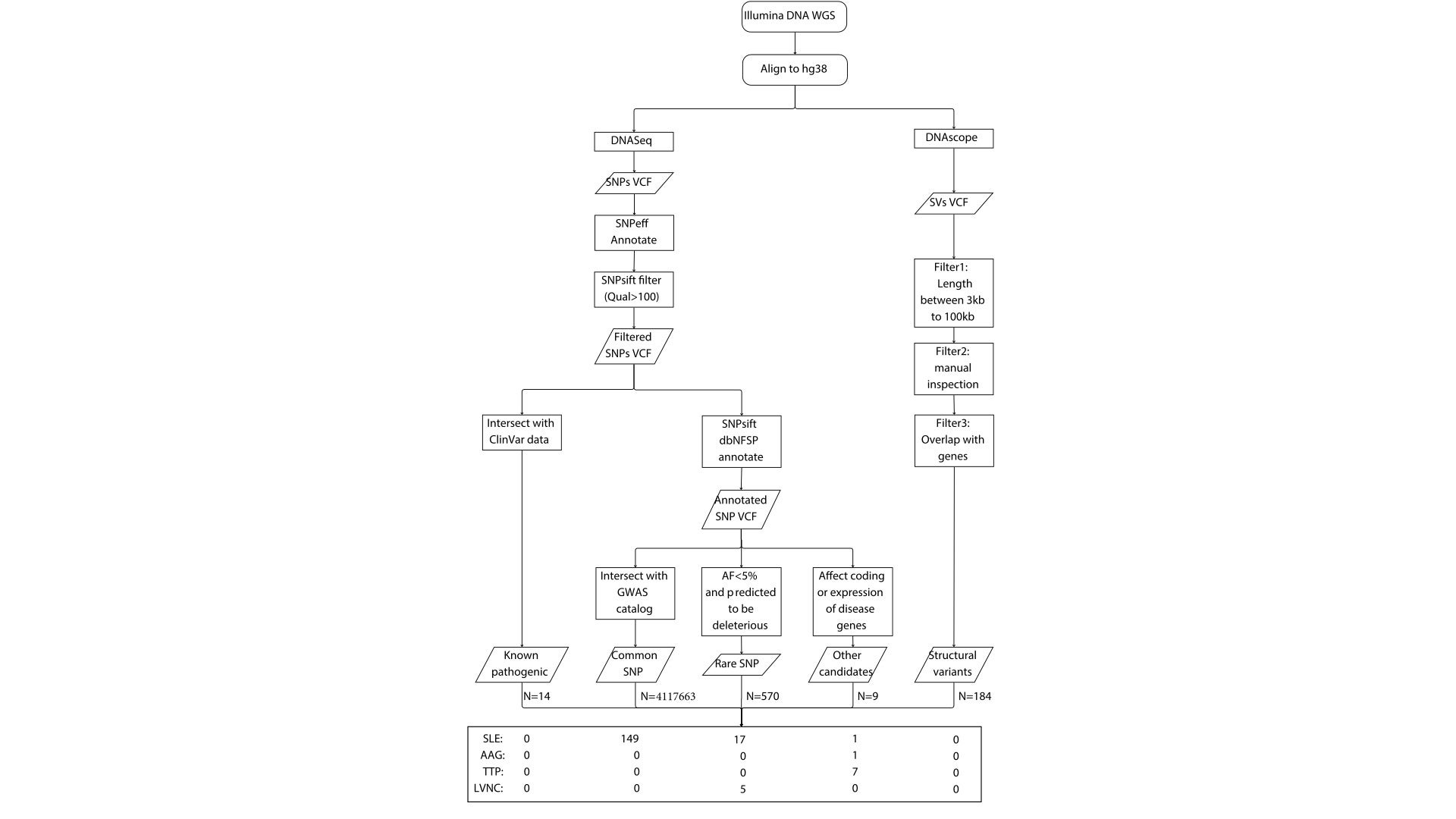
Figure 1. The analytic workflow to identify, annotate, and filter for the pathogenic genetic variants, including single-nucleotide polymorphisms (SNPs) and structural variants (SVs), for the four disease conditions. SLE: Systemic lupus erythematosus; AAG: Autoimmune autonomic ganglionopathy; TTP: Thrombotic thrombocytopenic purpura; LVNC: Left ventricular non-compaction.
| CHR | POS | ID | ALT | GT | P_VALUE | REPORTED_GENE |
|---|---|---|---|---|---|---|
| chr2 | 191071078 | rs10168266 | T | 0/1 | 1.00E-160 | STAT4 |
| chr7 | 128954129 | rs10488631 | C | 0/1 | 2.00E-143 | IRF5_-_TNPO3 |
| chr1 | 113834946 | rs2476601 | G | 1/1 | 4.00E-98 | PTPN22__AP4B1-AS1 |
| chr7 | 128933913 | rs4728142 | A | 0/1 | 7.00E-96 | KCP_-_IRF5 |
| chr8 | 11491677 | rs13277113 | A | 0/1 | 1.00E-88 | BLK |
| chr1 | 173257056 | rs704840 | G | 0/1 | 1.00E-46 | TNFSF4 |
| chr6 | 34752016 | rs9469857 | T | 0/1 | 9.00E-45 | RPS10P13_-_SNRPC |
| chr22 | 21585386 | rs5754217 | T | 0/1 | 7.00E-40 | UBE2L3 |
| chr5 | 160452971 | rs2431697 | C | 0/1 | 3.00E-37 | MIR3142HG |
| chr6 | 106120159 | rs548234 | T | 1/1 | 1.00E-35 | ATG5 |
| chr7 | 129089074 | rs2084654 | G | 1/1 | 8.00E-35 | TPI1P2_-_CYCSP20 |
| chr19 | 10364976 | rs2304256 | A | 1/1 | 9.00E-35 | TYK2 |
| chr2 | 191108837 | rs6752770 | G | 0/1 | 3.00E-34 | STAT4 |
| chr3 | 119431242 | rs3732421 | G | 0/1 | 5.00E-32 | TMEM39A |
| chr12 | 111569952 | rs653178 | T | 0/1 | 1.00E-31 | ATXN2 |
| chr2 | 191022906 | rs16833177 | C | 0/1 | 2.00E-31 | STAT4-AS1 |
| chr16 | 11100073 | rs3893660 | G | 0/1 | 3.00E-31 | CLEC16A |
| chr1 | 173376445 | rs10753071 | A | 1/1 | 4.00E-28 | TNFSF4 |
| chr8 | 11486171 | rs2254546 | G | 1/1 | 4.00E-28 | FAM167A_-_BLK |
| chr3 | 160028076 | rs485499 | C | 0/1 | 5.00E-25 | LINC01100__IL12A-AS1 |
| chr10 | 62045858 | rs4948496 | C | 0/1 | 2.00E-24 | ARID5B |
| chr10 | 48837411 | rs7922169 | G | 0/1 | 1.00E-23 | WDFY4 |
| chr17 | 39895095 | rs8067378 | G | 0/1 | 5.00E-23 | ZPBP2_-_GSDMB |
| chr19 | 10334150 | rs2278442 | A | 1/1 | 3.00E-22 | ICAM3 |
| chr2 | 203843923 | rs231804 | T | 0/1 | 1.00E-20 | RNU6-474P_-_CTLA4 |
| chr6 | 137825911 | rs670369 | T | 1/1 | 3.00E-20 | WAKMAR2 |
| chr6 | 25412759 | rs927984 | C | 1/1 | 1.00E-19 | CARMIL1__CMAHP |
| chr6 | 35149622 | rs1886243 | C | 0/1 | 3.00E-19 | TCP11_-_SCUBE3 |
| chr4 | 101816093 | rs10028805 | A | 1/1 | 4.00E-19 | BANK1 |
| chr11 | 35067136 | rs2732547 | C | 0/1 | 2.00E-18 | PDHX_-_CD44-DT |
| chr3 | 119399949 | rs11712165 | G | 0/1 | 5.00E-18 | ARHGAP31 |
| chr6 | 137806067 | rs675640 | C | 1/1 | 6.00E-18 | LINC02539_-_WAKMAR2 |
| chr2 | 30225478 | rs906868 | T | 0/1 | 1.00E-17 | H3P5_-_LBH |
| chr2 | 162254026 | rs2111485 | G | 0/1 | 2.00E-17 | FAP_-_IFIH1 |
| chr16 | 85985027 | rs2280381 | T | 0/1 | 4.00E-17 | LINC02132_-_LINC01082 |
| chr1 | 67354511 | rs6679356 | T | 1/1 | 8.00E-17 | IL12RB2 |
| chr8 | 10332530 | rs7832708 | T | 0/1 | 1.00E-16 | MSRA |
| chr8 | 8252414 | rs2945230 | G | 0/1 | 1.00E-16 | FAM86B3P_-_PRAG1 |
| chr1 | 192567683 | rs2816316 | A | 1/1 | 2.00E-16 | RGS21_-_RGS1 |
| chr1 | 67373890 | rs3828069 | C | 0/1 | 2.00E-16 | IL12RB2 |
| chr15 | 38541832 | rs8032939 | C | 1/1 | 4.00E-16 | RASGRP1 |
| chr4 | 971991 | rs11724804 | A | 0/1 | 4.00E-16 | DGKQ |
| chr18 | 69867948 | rs1790588 | C | 0/1 | 7.00E-16 | CD226 |
| chr3 | 58384450 | rs6445975 | T | 0/1 | 6.00E-15 | PXK |
| chr8 | 11514632 | rs11250141 | A | 1/1 | 6.00E-15 | BLK |
| chr6 | 25428726 | rs722086 | T | 1/1 | 7.00E-15 | CARMIL1__CMAHP |
| chr2 | 203768433 | rs10932020 | A | 0/1 | 1.00E-14 | KRT18P39_-_NPM1P33 |
| chr3 | 28034494 | rs427221 | C | 0/1 | 1.00E-14 | LINC01967 |
| chr4 | 101874688 | rs6816787 | A | 0/1 | 2.00E-14 | BANK1 |
| chr11 | 118638950 | rs483598 | G | 1/1 | 4.00E-14 | PHLDB1 |
| chr11 | 641563 | rs11246228 | C | 0/1 | 4.00E-14 | DRD4_-_DEAF1 |
| chr19 | 10491504 | rs11085735 | C | 1/1 | 4.00E-14 | KEAP1 |
| chr6 | 25683052 | rs1883259 | C | 0/1 | 1.00E-13 | SCGN |
| chr16 | 58219556 | rs2731783 | G | 1/1 | 2.00E-13 | RN7SL645P_-_CFAP263 |
| chr17 | 75330884 | rs4542691 | C | 0/1 | 2.00E-13 | GRB2 |
| chr4 | 102636062 | rs2866414 | T | 0/1 | 2.00E-13 | MANBA |
| chr7 | 129101859 | rs4731549 | T | 0/1 | 2.00E-13 | TPI1P2_-_CYCSP20 |
| chr6 | 90170674 | rs10944479 | A | 0/1 | 5.00E-13 | BACH2 |
| chr6 | 106135045 | rs742108 | A | 0/1 | 9.00E-13 | ATG5 |
| chr1 | 183412660 | rs11577151 | A | 0/1 | 1.00E-12 | NMNAT2 |
| chr5 | 100793209 | rs4703116 | C | 0/1 | 1.00E-12 | RN7SKP62_-_ST8SIA4 |
| chr11 | 128479083 | rs10790956 | G | 0/1 | 2.00E-12 | ETS1 |
| chr1 | 183291524 | rs946173 | G | 0/1 | 2.00E-12 | NMNAT2 |
| chr19 | 10289228 | rs2569693 | T | 0/1 | 2.00E-12 | LIMASI__ICAM4-AS1 |
| chr5 | 134084952 | rs244673 | A | 1/1 | 2.00E-12 | VDAC1_-_TCF7 |
| chr5 | 151097526 | rs4958890 | T | 0/1 | 2.00E-12 | TNIP1_-_ANXA6 |
| chr6 | 25787589 | rs2096386 | A | 0/1 | 2.00E-12 | SLC17A1 |
| chr8 | 10728173 | rs7836366 | G | 0/1 | 2.00E-12 | PINX1__SOX7 |
| chr11 | 64360951 | rs516124 | T | 0/1 | 3.00E-12 | RPS6KA4 |
| chr11 | 65775268 | rs489574 | A | 0/1 | 3.00E-12 | AP5B1 |
| chr2 | 102473668 | rs17027255 | T | 0/1 | 3.00E-12 | SLC9A4 |
| chr22 | 39325740 | rs12627970 | G | 0/1 | 4.00E-12 | RPL3_-_SYNGR1 |
| chr1 | 42051099 | rs2042065 | C | 1/1 | 5.00E-12 | HNRNPFP1_-_GUCA2B |
| chr6 | 137878280 | rs610604 | T | 0/1 | 5.00E-12 | TNFAIP3 |
| chr6 | 34936315 | rs847852 | A | 0/1 | 6.00E-12 | ANKS1A |
| chr1 | 161492938 | rs4657039 | G | 0/1 | 7.00E-12 | RNU6-481P_-_FCGR2A |
| chr18 | 12797695 | rs2847297 | G | 0/1 | 7.00E-12 | PTPN2 |
| chr1 | 92469854 | rs6662618 | G | 1/1 | 1.00E-11 | RN7SL824P_-_GFI1 |
| chr1 | 173197108 | rs1234313 | G | 0/1 | 2.00E-11 | TNFSF4 |
| chr3 | 159783884 | rs6441275 | A | 1/1 | 2.00E-11 | SCHIP1__IQCJ-SCHIP1 |
| chr14 | 102797451 | rs12148050 | G | 1/1 | 3.00E-11 | TRAF3 |
| chr3 | 58346341 | rs4681679 | G | 1/1 | 3.00E-11 | PXK |
| chr6 | 90247744 | rs3757247 | T | 1/1 | 4.00E-11 | BACH2 |
| chr6 | 106136013 | rs12213031 | T | 0/1 | 5.00E-11 | ATG5 |
| chr8 | 42283286 | rs4560769 | G | 0/1 | 5.00E-11 | IKBKB |
| chr9 | 120927961 | rs3761847 | A | 0/1 | 5.00E-11 | TRAF1 |
| chr10 | 48768184 | rs17836440 | C | 0/1 | 6.00E-11 | WDFY4 |
| chr17 | 39354149 | rs9892055 | T | 0/1 | 6.00E-11 | FBXL20 |
| chr6 | 25413694 | rs1543603 | G | 1/1 | 6.00E-11 | CARMIL1__CMAHP |
| chr3 | 137049168 | rs17374749 | A | 0/1 | 8.00E-11 | IL20RB_-_RNA5SP142 |
| chr1 | 67361470 | rs6685236 | A | 1/1 | 1.00E-10 | IL12RB2 |
| chr2 | 100142995 | rs11681966 | A | 0/1 | 1.00E-10 | AFF3 |
| chr2 | 190391157 | rs11892526 | T | 1/1 | 1.00E-10 | INPP1_-_MFSD6 |
| chr2 | 190754098 | rs4853732 | T | 0/1 | 1.00E-10 | NAB1_-_GLS |
| chr16 | 11260640 | rs243325 | C | 1/1 | 2.00E-10 | RMI2 |
| chr2 | 65365191 | rs6738087 | C | 1/1 | 2.00E-10 | SPRED2 |
| chr6 | 25684177 | rs4711092 | C | 1/1 | 2.00E-10 | SCGN |
| chr8 | 11753534 | rs804283 | A | 0/1 | 2.00E-10 | GATA4 |
| chr2 | 134293051 | rs13014122 | A | 0/1 | 6.00E-10 | MGAT5 |
| chr3 | 119410956 | rs11714363 | A | 0/1 | 6.00E-10 | ARHGAP31 |
| chr19 | 10458967 | rs2358681 | C | 1/1 | 7.00E-10 | PDE4A |
| chr3 | 121834872 | rs12490565 | A | 0/1 | 8.00E-10 | IQCB1 |
| chr3 | 12218116 | rs310746 | T | 1/1 | 8.00E-10 | SYN2_-_GSTM5P1 |
| chr1 | 198687278 | rs2359952 | G | 0/1 | 1.00E-09 | PTPRC |
| chr2 | 190316755 | rs291458 | C | 0/1 | 1.00E-09 | HIBCH |
| chr6 | 25437758 | rs6900224 | T | 0/1 | 1.00E-09 | CARMIL1__CMAHP |
| chr12 | 120818717 | rs3883901 | C | 0/1 | 2.00E-09 | SPPL3 |
| chr6 | 137697302 | rs6917441 | G | 0/1 | 2.00E-09 | LINC03004 |
| chr7 | 28145472 | rs849141 | G | 0/1 | 2.00E-09 | JAZF1 |
| chr8 | 128426805 | rs6470627 | G | 1/1 | 2.00E-09 | LINC00824 |
| chr16 | 11269835 | rs415595 | G | 0/1 | 3.00E-09 | RMI2 |
| chr16 | 57389946 | rs8063316 | T | 1/1 | 3.00E-09 | CX3CL1_-_CCL17 |
| chr8 | 8957380 | rs11778913 | A | 0/1 | 3.00E-09 | RNU6-682P_-_SNORA70 |
| chr10 | 62664778 | rs10995263 | T | 0/1 | 4.00E-09 | LINC02929 |
| chr11 | 128416471 | rs1547892 | C | 0/1 | 4.00E-09 | LINC02098_-_ETS1 |
| chr1 | 235876577 | rs9782955 | C | 0/1 | 4.00E-09 | LYST |
| chr2 | 190887601 | rs1921915 | A | 1/1 | 4.00E-09 | GLS |
| chr7 | 50209417 | rs11761913 | T | 0/1 | 4.00E-09 | SPMIP7_-_IKZF1 |
| chr8 | 9109096 | rs17155242 | G | 0/1 | 4.00E-09 | ERI1 |
| chr17 | 7323638 | rs35776863 | A | 0/1 | 5.00E-09 | NEURL4 |
| chr6 | 36655123 | rs1321311 | A | 0/1 | 5.00E-09 | RNU1-88P_-_Y_RNA |
| chr5 | 160470788 | rs1862390 | T | 1/1 | 6.00E-09 | MIR3142HG |
| chr12 | 111489935 | rs7134740 | A | 0/1 | 7.00E-09 | ATXN2 |
| chr20 | 59159698 | rs1535271 | A | 0/1 | 7.00E-09 | ZNF831 |
| chr5 | 56148813 | rs12515884 | C | 0/1 | 7.00E-09 | ANKRD55 |
| chr17 | 75420733 | rs9913538 | T | 0/1 | 8.00E-09 | RNU6-938P_-_Y_RNA |
| chr1 | 8422018 | rs301806 | T | 0/1 | 8.00E-09 | RERE |
| chr8 | 8901751 | rs332013 | C | 0/1 | 8.00E-09 | RNU6-682P_-_SNORA70 |
| chr10 | 62296309 | rs1835862 | A | 1/1 | 9.00E-09 | LINC02621 |
| chr1 | 183816708 | rs2500125 | C | 1/1 | 9.00E-09 | RGL1 |
| chr6 | 25903080 | rs16891146 | A | 0/1 | 9.00E-09 | H2BP5_-_SLC17A2 |
| chr11 | 65784486 | rs479844 | G | 0/1 | 1.00E-08 | AP5B1_-_OVOL1 |
| chr1 | 196693329 | rs10922096 | C | 0/1 | 1.00E-08 | CFH |
| chr2 | 203866221 | rs733618 | C | 0/1 | 1.00E-08 | CTLA4 |
| chr5 | 151109829 | rs1133202 | G | 0/1 | 1.00E-08 | ANXA6 |
| chr6 | 25684051 | rs4712955 | G | 0/1 | 1.00E-08 | SCGN |
| chr2 | 213011526 | rs12470231 | G | 0/1 | 2.00E-08 | IKZF2 |
| chr4 | 122631659 | rs2893008 | G | 0/1 | 2.00E-08 | IL21-AS1 |
| chr5 | 159288655 | rs4921458 | C | 0/1 | 2.00E-08 | UBLCP1_-_IL12B |
| chr10 | 12445997 | rs6602588 | A | 0/1 | 3.00E-08 | CAMK1D |
| chr1 | 183207295 | rs525410 | G | 0/1 | 3.00E-08 | LAMC2 |
| chr6 | 129873059 | rs6939565 | T | 1/1 | 3.00E-08 | TMEM244_-_L3MBTL3 |
| chr7 | 128785714 | rs339083 | T | 0/1 | 3.00E-08 | OPN1SW_-_CCDC136 |
| chr6 | 36382113 | rs879036 | C | 0/1 | 4.00E-08 | ETV7 |
| chr8 | 11246386 | rs958648 | G | 1/1 | 4.00E-08 | LINC00529 |
| chr10 | 5844771 | rs2380205 | T | 0/1 | 5.00E-08 | GDI2_-_ANKRD16 |
| chr15 | 75071016 | rs1867147 | T | 0/1 | 5.00E-08 | PPCDC |
| chr2 | 191220415 | rs6434455 | G | 1/1 | 5.00E-08 | MYO1B-AS1 |
| chr3 | 159992603 | rs583911 | A | 0/1 | 5.00E-08 | IL12A__IL12A-AS1 |
Table 1. 149 signature single-nucleotide polymorphisms with known association with systemic lupus erythematosus carried by the patients.
Abbreviations: CHR, chromosome; POS, position; ALT, alternative allele; GT, genotype. 0/1: heterozygous alternative allele; 1/1: homozygous alternative allele.
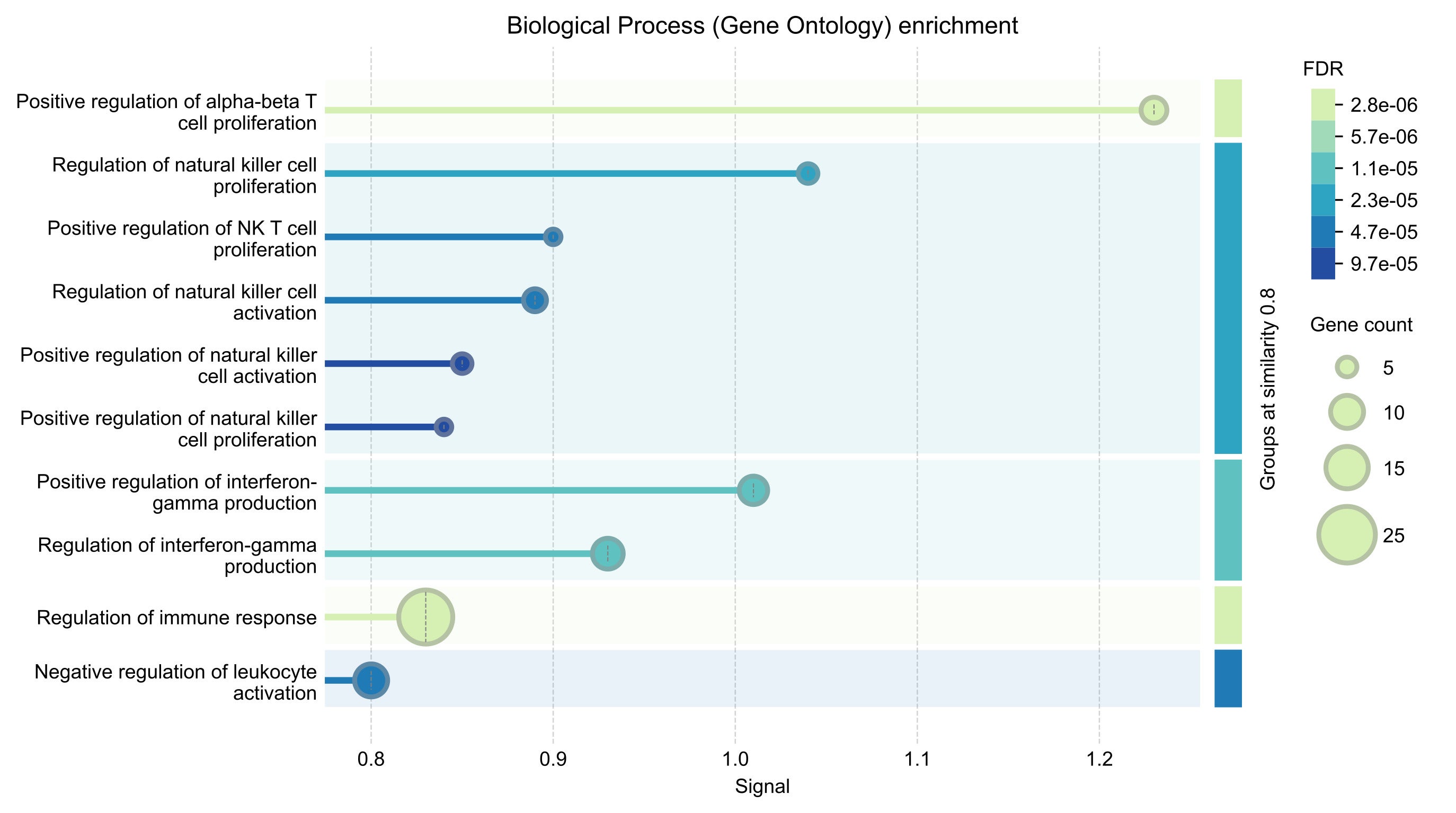
Figure 2. The 120 protein-coding genes mapped by the signature single-nucleotide polymorphisms with known association with systemic lupus erythematosus are enriched in the immune response processes. The level of enrichment is measured by the signal, which is a weighted harmonic mean between the observed/expected ratio and the negative-log transformed false discovery rate (FDR). The biological process categories were grouped by similarity (shared genes > 0.8)
We further documented a total of 535 SNPs that were significantly associated with SLE in a total of 74 GWAS reports34 (Supplementary File 3). Notably, the significance of the associations, indicated by the P-values, depends on cohort sizes and ancestries and should, therefore, not be compared across different studies. In addition, the list includes SNPs in large gene families such as the mucin genes (e.g., MUC21) and the human leukocyte antigen (HLA) genes (e.g., HLA-DRA). The standard Illumina sequencing technology, however, generates short sequencing reads that are prone to misalignment across different gene paralogs, resulting in a large number of false positive SNPs. Despite being one of the most significantly associated loci for SLE35–36, further tests are required to validate the identified SNPs in the complex region of HLA genes.
Unlike SLE, there are currently no systematic association studies for AAG, TTP, and LVNC due to their rareness. Thus, we did not report any common SNP that could be associated with these three diseases.
Rare, likely pathogenic genetic variants in disease-associated genes
Based on ten in silico prediction algorithms of the SNP effects, we report 570 rare, likely deleterious SNPs in the patient (Supplementary File 4). Five genes carried >=3 of such mutations, including C6orf10(N=3), DNAH1(N=3), HLA-DQB1(N=6), LILRA2(N=3), and USH2A(N=3). Gene C6orf10, also known as TSBP1, has been associated with autoimmune diseases, including SLE, in European37 and East Asian38 populations. Mutations in HLA-DQB1 will need independent validations. DNAH1, LILRA2, and USH2A have no known associations with the reported diseases. USH2A, for instance, is one of the largest genes and tends to contain more mutations. A sixth gene, PABPC3 (N=12*)*, was filtered as the mutations were likely alignment errors and failed on the manual inspection.
Most rare, likely deleterious SNPs affect the same genes once or twice. Using a list of 525 SLE-associated genes based on literature curation39–40, we identified 17 genes with likely functional perturbations from rare SNPs, predicted by a minimum of two methods (Table 2). The two genes with the highest strength of functional associations are DNASE1 (rs1799891) and IL21R (rs146177564). DNASE1 encodes the enzyme DNase 1, which is localized in the zymogen granules of the nuclear envelope to break down DNA in a manner known as endonucleolytic cleavage41. Anti-nuclear antibodies against naked DNA and intact nucleosomes are hallmarks of SLEs42. A loss-of-function mutation in the DNASE1 gene could theoretically cause inadequate clearance of chromatin or chromatin-protein complexes following cellular apoptosis, therefore triggering the autoimmune responses. However, an in vitro assay found that rs1799891 is likely a gain-of-function mutation that upregulates the DNase 1 activity43. Thus, until we can measure the IgG titer of dsDNA and nucleosomal antigens in the patients, the effect of the DNASE1 mutation, rs1799891, on SLE symptoms remains inconclusive.
IL21R encodes the cytokine receptor for interleukin 21 (IL-21) and is another known susceptibility gene for SLE44. IL-21 is a pleiotropic cytokine with multiple effects on the immune system. Its receptor, IL-21R, is widely expressed in B cells, T cells, and natural killer cells but is underexpressed in SLE patients. This reduced expression has been significantly linked to elevated levels of anti-ds DNA antibodies and B lymphocytopenia (a decrease in B cells)45.
| CHR | POS | ID | ALT | GT | GENE | TYPE | #METHODa | STRENGTHb |
|---|---|---|---|---|---|---|---|---|
| chr16 | 3657022 | rs1799891 | G | 0/1 | DNASE1(Pro154Ala) | missense | 5 | 2.88 |
| chr16 | 27443024 | rs146177564 | T | 0/1 | IL21R(Arg161Cys) | missense | 3 | 2.88 |
| chr7 | 87531302 | rs2032582 | T | 0/1 | ABCB1(Ser893Thr) | missense | 5 | 1.656 |
| chr6 | 31816086 | rs562047 | C | 1/1 | HSPA1A(Glu110Asp) | missense | 2 | 1.591 |
| chr13 | 95210754 | rs11568658 | A | 0/1 | ABCC4(Gly187Trp) | missense | 8 | 1.306 |
| chr9 | 122381694 | rs5789 | A | 0/1 | PTGS1(Leu237Met) | missense | 5 | 1.293 |
| chr10 | 128105328 | rs45549235 | A | 0/1 | MKI67(Ala2171Val) | missense | 2 | 1.267 |
| chr6 | 121447661 | rs376074787 | C | 0/1 | GJA1(Ser272Pro) | missense | 5 | 1.246 |
| chr8 | 11845196 | rs79487342 | T | 0/1 | CTSB(Asp317Asn) | missense | 3 | 1.238 |
| chr15 | 29732685 | rs759848653 | T | 0/1 | TJP1(Ala690Thr) | missense | 4 | 1.214 |
| chr10 | 73915328 | rs72816325 | C | 0/1 | PLAU(Tyr350His) | missense | 6 | 1.2 |
| chr6 | 41936044 | rs1051130 | C | 0/1 | CCND3(Ser259Ala) | missense | 2 | 1.156 |
| chr20 | 391432 | rs41281850 | A | 0/1 | TRIB3(Ser173Asn) | missense | 7 | 1.123 |
| chr17 | 82091449 | rs759242677 | T | 0/1 | FASN | structural_interaction | 2 | 1.101 |
| chr7 | 100893176 | rs1799805 | T | 0/1 | ACHE(His353Asn) | missense | 3 | 1.07 |
| chr5 | 169594617 | rs185497566 | C | 0/1 | SPDL1(Lys276Thr) | missense | 6 | 1.029 |
| chr1 | 181089755 | rs3747951 | T | 0/1 | IER5(Pro285Ser) | missense | 5 | 1.01 |
Table 2. 17 genes with rare, likely pathogenic mutations for systemic lupus erythematosus.
Abbreviations: CHR, chromosome; POS, position; ALT, alternative allele; GT, genotype. 0/1: heterozygous alternative allele; 1/1: homozygous alternative allele.
a indicates the number of methods that predict the SNP to be deleterious.
b indicates the relative strength of functional association between the gene and Systemic Lupus Erythematosus.
Using a similar approach, we curated a list of 78 LVNC-associated genes based on a systematic review, including 561 patients from 17 studies16. The list of genes includes those related to sarcomeres (the basic contractile unit in muscle), arrhythmias (abnormal heartbeat), and mitochondrial functions, among others. The patient carries rare, deleterious SNPs in 5 of the 78 genes, including sarcomere genes MYBPC3, TNNT1, and OBSCN, arrhythmia gene ANK2, and a non-sarcomere non*-*arrhythmia gene PLEC (Table 3). Notably, OBSCN, ANK2, PLEC and especially TNNT1 are long genes, which may increase the likelihood of SNPs. Nevertheless, observing multiple gene mutations associated with LVNC suggests a significant genetic risk from rare SNPs. Among these, MYBPC3 encodes the cardiac isoform of myosin-binding protein and is exclusively expressed in cardiac muscle, where it plays a crucial role in regulating cardiac contraction46. TNNT1 (also called TNT) encodes a large, abundant protein of striated muscle termed titin, and its truncation mutations are associated with a higher risk of heart failure among LVNC patients47. The patient instead carries rs112562759 in TNNT1, a missense/splice variant, and the impact is expected to be lower than truncations. OBSCN encodes obscurin that interacts with titin and is involved in sarcomere assembly and development48. ANK2 is highly expressed in the heart, and its protein is crucial for localizing and stabilizing ion transporters and ion channels in cardiomyocytes49. Individuals carrying ANK2 variants displayed an increased maximum wall thickness in the left ventricle, which is characteristic of hypertrophic cardiomyopathy50. Finally, PLEC encodes plectin, which links intermediate filaments (IFs) to Z-discs in sarcomeres and is essential for the proper cytoarchitecture during heart development51. The same filtering criteria did not identify any rare, deleterious mutations in the risk genes, such as CHRNA3 for AAG or ADAMTS13 for TTP.
| CHR | POS | ID | ALT | GT | GENE | TYPE | #METHODa |
|---|---|---|---|---|---|---|---|
| chr1 | 228372266 | rs72762066 | T | 0/1 | OBSCN(Pro8120Leu) | missense | 2 |
| chr4 | 113369549 | rs150808807 | G | 0/1 | ANK2(Gln3785Arg) | missense | 2 |
| chr8 | 143930279 | rs369344419 | C | 0/1 | PLEC(Asp963Gly) | missense | 8 |
| chr11 | 47333924 | rs11570112 | C | 0/1 | MYBPC3(Gln998Glu) | missense&splice | 4 |
| chr19 | 55147019 | rs112562759 | C | 0/1 | TNNT1(Glu12Gly) | missense&splice | 5 |
Table 3. 5 genes with rare, likely pathogenic mutations for left ventricular non-compaction.
Abbreviations: CHR, chromosome; POS, position; ALT, alternative allele; GT, genotype. 0/1: heterozygous alternative allele; 1/1: homozygous alternative allele.
a indicates the number of methods that predict the SNP to be deleterious
Other candidate genetic variants in disease-associated genes
Despite our usage of 11 in silico methods, their combined power to predict SNP effects may cause under-reporting of pathogenic variants, especially for rare disorders. Thus, we reported 7 additional candidate pathogenic variants in the genes with known risks for SLE, AAG, and TTP (Table 4). Of these, rs45487196 is a rare, missense mutation (p.Val187Ile) in gene C1QC, which encodes the C-chain polypeptide of the complement subcomponent C1q. Monogenic mutations that led to C1q deficiency are currently acknowledged as one of the most significant genetic factors associated with SLE5, 28. The lack of C1q leads to poor clearance of dead cells, leading to intracellular molecules releasing and exposing, thus triggering the development of autoantibodies, as described in the Waste Disposal Theory for SLE52. In addition, C1q can significantly activate DNase I in the clearance of chromatin derived from necrotic cells53. Thus, despite rs45487196 being characterized as benign or tolerable for C1QC, it remains a possible pathogenic mutation through interactions or additive effects to the DNASE1 mutation (Table 3).
| CHR | POS | ID | ALT | GT | GENE | TYPE | DISEASE |
|---|---|---|---|---|---|---|---|
| chr1 | 22647604 | rs45487196 | A (Val187Ile) | 0/1 | C1QC | missense | SLE |
| chr15 | 78601522 | rs141141954 | T (Gly374Ser) | 0/1 | CHRNA3 | missense | AAG |
| chr9 | 133447776 | rs655911 | C | 1/1 | ADAMTS13 | intronic | TTP |
| chr9 | 133460340 | rs2301615 | T | 1/1 | ADAMTS13 | downstream | TTP |
| chr9 | 133460785 | rs1858879 | C | 1/1 | ADAMTS13 | downstream | TTP |
| chr9 | 133466757 | rs28660186 | C | 1/1 | ADAMTS13 | downstream | TTP |
| chr9 | 133474675 | rs4962155 | A | 1/1 | ADAMTS13 | downstream | TTP |
Table 4. Additional single-nucleotide polymorphisms associated with systemic lupus erythematosus (SLE), Autoimmune autonomic ganglionopathy (AAG), and Thrombotic thrombocytopenic purpura (TTP).
Abbreviations: CHR, chromosome; POS, position; ALT, alternative allele; GT, genotype. 0/1: heterozygous alternative allele; 1/1: homozygous alternative allele.
In addition, the patient carries a rare, missense mutation in CHRNA3, rs141141954 (p. Gly374Ser), which is predicted to have a low impact on the protein function (Table 4). CHRNA3 encodes the neuronal acetylcholine receptor subunit alpha-3 (nAChRα3), which is essential for ganglionic synaptic transmission and is the only known gene that increases genetic risk for AAG. Based on two familial AAG families, within which the disease was transmitted across generations, a frameshift mutation (p. Leu303Aspfs*115) and a missense mutation (p. Asp230Asn) in CHRNA3 were reported as the causal mutations8. However, no other family members of the patient of our study have AAG. The mutated amino acid is also predicted to be intracellular54 and inaccessible by serum antibodies (Figure 3). Thus, unless rs141141954 is a de novo mutation unique to the patient and the intracellular mutation could affect the extracellular protein structures, rs141141954 likely had not played a causal role in the AAG disease.
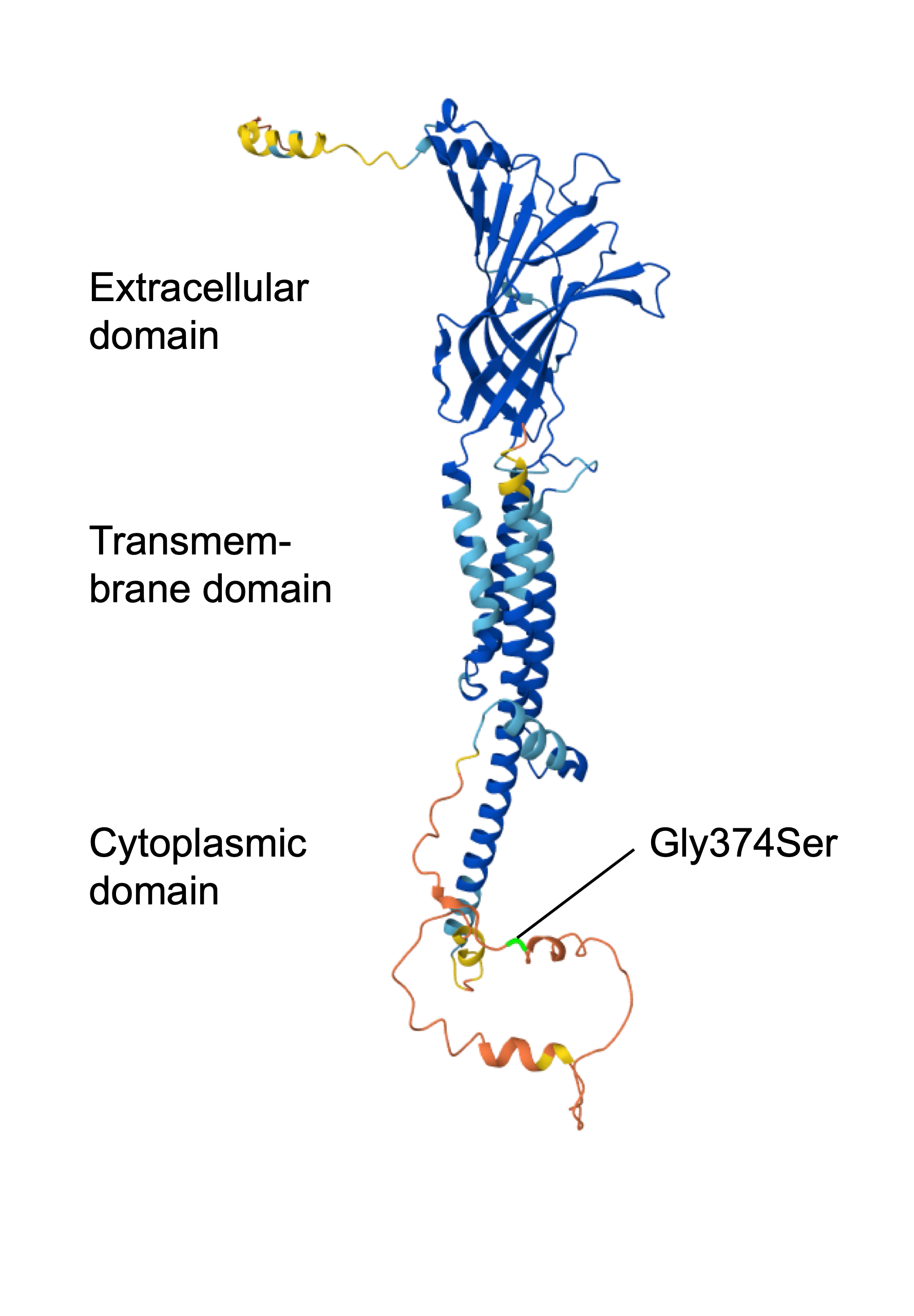
Figure 3. The predicted protein structure of the neuronal acetylcholine receptor subunit alpha-3, a protein associated with autoimmune autonomic ganglionopathy. The patient carries a missense mutation, Gly374Ser, which is predicted to be in the cytoplasmic domain.
Biallelic (homozygous or compound heterozygous) pathogenic variants in the gene ADAMTS13 cause familial TTP, a rare form of TTP55. The patient carries no exonic SNPs in ADAMTS13 but has five homozygous intronic or intergenic SNPs in the regulatory regions that could decrease the gene expression. Human endothelial cells from the blood vessels synthesize and release functional ADAMTS13 proteins into the blood. We searched for intronic or intergenic SNPs in the c (eQTLs) predicted to downregulate the ADAMTS13 transcription levels in the aorta or tibial arteries32. Out of a total of 38 downregulating SNPs, the patient carries seven, 2 heterozygous and 5 homozygous, SNPs that could potentially reduce the expression of ADAMTS13 in the source cells and, in turn, decrease the ADAMTS13 protein in the blood (Table 4).
Discussion
In this study, we utilized state-of-the-art computational tools for analyzing the genomic sequence of a patient with a rare combination of diseases, including SLE, AAG, TTP, and LVNC. After applying thorough filters to eliminate noise and conducting comprehensive literature reviews of known genetic factors, we identified likely pathogenic mutations in the patient’s genome across various categories. These include a significant number of common mutations shared with others that pose modest, predisposing risks, as well as several rare mutations that are unique to the patient and likely have a greater impact on their diseases. As expected, the majority of the mutations are linked to conditions that have a well-established genetic basis, such as SLE and LVNC. Notably, the list of reported genetic risks may be biased towards autosomes (i.e., non-sex chromosomes). This is likely due to standard genotyping methods, such as the SNP arrays used in previous GWAS analyses and the short-read DNA sequencing employed in this study, which can produce false mutations due to sequence homology between the X and Y chromosomes.
One of the other main challenges in identifying the credible genetic factors in the patient lies in the rarity of her disease. Globally, approximately 7,000 rare diseases, defined as fewer than 1 in 15,000 individuals in the United States or under 1 in 2,000 in Europe, have been identified thus far. While each rare disease affects a small segment of the population, together, approximately 350 million people worldwide are impacted, leading to life-threatening challenges or chronic disabilities. It is estimated that around 80% of these diseases have a genetic basis56. However, most patients remain without a genetic diagnosis. The symptoms of rare diseases frequently mimic those of more prevalent conditions, complicating the diagnostic process. Additionally, these diseases can exhibit highly individualized characteristics influenced by a combination of genetic factors and environmental conditions.
Creating direct collaborations and sharing information between patients, physicians, and researchers is crucial for addressing the specific challenges associated with rare or complex diseases. Our analysis of the presented case is limited by the current understanding of the diseases’ etiology and its interactions. However, we believe that the study model, which promotes multidisciplinary collaboration and includes a direct partnership with the patient, will encourage similar case studies in the future. This approach will help accumulate more cases that can be cross-referenced, addressing the challenge posed by the limited occurrence of rare diseases. As sequencing costs continue to decrease and genomic data expands, this study model will also enhance research accessibility for patients.
Acknowledgments
This work utilized computing resources provided by the Stanford Genetics Bioinformatics Service Center and the City University of Hong Kong high-performance computing (HPC) resources.
Funding: This work was supported by the City University of Hong Kong New Faculty Fund 9610590.
Conflict of Interest
The authors have no relevant financial or non-financial interests to disclose.
Code Availability
The code used in this study is in Supplementary File 5.
Supplementary Information
Methods
Whole genome sequencing and genetic variant calling
DNA extracted from the patient’s saliva sample was used for whole genome sequencing on the Illumina sequencing platform for an average sequencing depth of 51×. The sequencing reads were aligned to the human reference genome hg38 containing alternative and decoy sequences (http://ftp.1000genomes.ebi.ac.uk/vol1/ftp/technical/reference/GRCh38_reference_genome/GRCh38_full_analysis_set_plus_decoy_hla.fa), using the alt-aware aligner BWA (v0.7.12)22. The germline single nucleotide polymorphisms and short insertions and deletions were identified by the Sentieon DNAseq pipeline (v202308.03) and the structural variants were called using the Sentieon DNAscope pipeline (v202308.03)23. The overall analytic workflow is summarized in Figure 1, and the code is in Supplementary File 5.
Annotation of the genetic variants
The basic function and effects of the detected genetic variants were first annotated using SnpEff(v4.3t)57. Next, SnpSift(v4.3t)58 was employed to further filter low-quality (<100) variant calls and annotate the variants, including (1) known clinically pathogenic or likely pathogenic mutations based on the ClinVar database27, (2) population allele frequencies of the alternative alleles based on the ExAC database59, (3) SNP effects with up to 11 in silico prediction algorithms as summarized by the dbNSFP database (v4.1)60, and (4) known SNPs with significant associations to SLE risk based on the genome-wide association studies catalog34.
Filtering for rare, likely pathogenic SNPs
Rare SNPs were defined as those with a global allele frequency below 5%, based on the ExAC database. The SNP effects were reported with 10 in silico prediction methods, including (1) CADD61, (2) SIFT62, (3) PROVEAN63, (4) MutationTaster64, (5) MetaSVM65, (6) LRT66, (7) FATHMM67 (8) Polyphen2_HVAR68, (9) Polyphen2_HDIV68, and (10) MutationAssessor69. A pathogenic SNP is defined if it was predicted to be intolerant or related to disease in at least two methods (Table 2 and Table 3).
Expression Quantitative Trait Loci for ADAMTS13
9,446 single tissue expression quantitative trait loci and tissue combinations that could significantly affect ADAMTS13 expression levels were obtained from the Genotype-Tissue Expression database32. We filtered for the homozygous SNPs that could downregulate ADAMTS13 expression, with a normalized effect size less than -0.2, in blood vessels (Aorta or Tibial).
Supplementary Figures, Tables, and Data
**All non-linked supplementary files are available by request only. Please email us at: journal@researchtothepeople.org to request access.
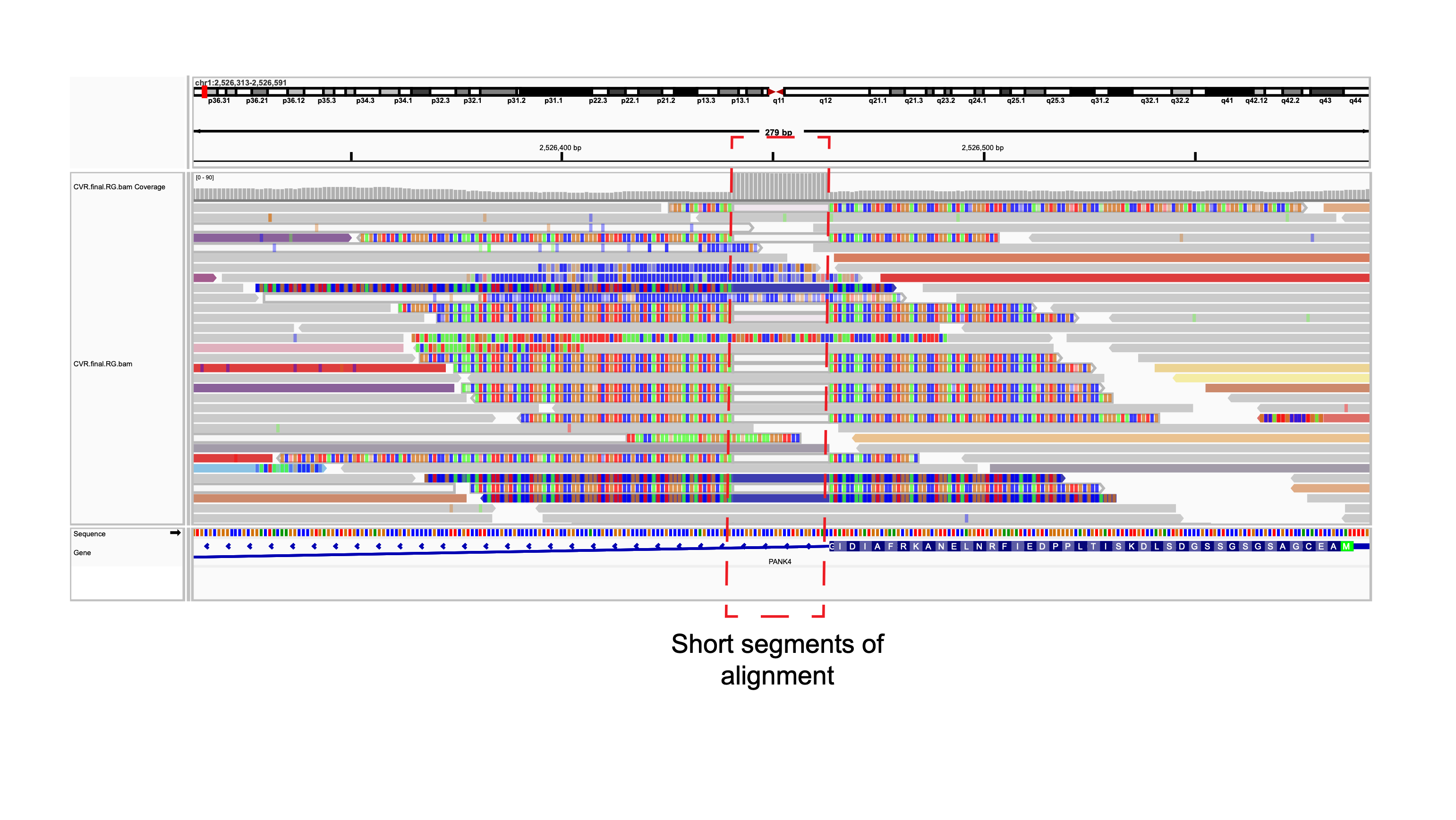
Figure S1. Misalignment of microorganism-derived sequencing reads leads to false insertion structural variations calls.
Supplementary Table 1. 14 pathogenic or likely pathogenic mutations annotated by the ClinVar database.
Supplementary Table 2. 184 structural variants between 3 and 100kb.
Supplementary Table 3. 28 genes affected by structural variants.
Supplementary File 1. All single nucleotide variants and short insert or deletions identified by the Sentieon DNAseq pipeline, with SNPeff and SNPsift annotation.
Supplementary File 2. All structural variants identified by the Sentieon DNAscope pipeline.
Supplementary File 3. 535 SNPs with significant associations to systemic lupus erythematosus.
Supplementary File 4. 570 rare, likely deleterious SNPs.
Supplementary File 5. Code used in the bioinformatics pipeline.
Be the first to comment on this publication.