Purpose
Pathogenic biallelic variants in ITGA3 cause a rare multisystem syndrome involving interstitial lung disease, nephropathy, and epidermolysis bullosa. However, the mechanisms linking impaired alpha-3 integrin function to progressive fibroinflammatory lung remodeling remain incompletely defined. Here, we integrated multi-omics to uncover the disease architecture in a patient with compound heterozygous ITGA3 variants and severe pulmonary fibrosis. Long-read RNA sequencing confirmed the trans expression of a splice-donor variant and a missense variant (p.Thr658Arg). Systemically, plasma proteomics and PBMC single-cell RNA-sequencing revealed disrupted cytoskeletal dynamics alongside an inflammatory, monocyte-skewed immune state with altered antigen presentation. At the tissue level, spatially resolved transcriptomics of longitudinal lung biopsies identified the expansion of a fibrotic damage-associated transient progenitor (DATP)-like transitional AT2 epithelial state. This population was notably enriched for epithelial-injury and aberrant-basaloid signatures. Spatial neighborhood analysis demonstrated an intensifying co-occurrence between these transitional AT2 cells and fibroblasts, revealing an active, localized epithelial-fibroblast remodeling niche. Collectively, this multi-omics framework captures a baseline of systemic immune and cytoskeletal dysregulation, culminating in the major finding of an active, DATP-like transitional AT2-fibroblast niche driving progressive lung remodeling.
Introduction
Pathogenic biallelic variants in ITGA3 (encoding integrin alpha-3) cause a rare, devastating multisystem syndrome characterized by interstitial lung disease, nephrotic syndrome, and epidermolysis bullosa1. As a critical basement-membrane adhesion receptor, integrin alpha-3 maintains epithelial integrity across the lung, kidney, and skin. In the respiratory tract, the disruption of these vital epithelial-basement-membrane interactions creates a profound vulnerability to recurrent injury and maladaptive repair. While the broad consequences of impaired integrin function are known to involve disrupted cytoskeletal organization, immune remodeling, and fibroblast activation2–3, the precise cellular mechanisms that propagate this localized tissue remodeling remain poorly characterized.
While genomic sequencing can successfully establish a molecular diagnosis of ITGA3-associated disease, variant interpretation alone fundamentally cannot resolve the spatial and cellular architecture of the resulting lung pathology. This gap between genetic diagnosis and tissue-level mechanism is especially critical in patients with complex, multisystem presentations, where interstitial fibrosis, constrictive bronchiolitis, systemic immune dysregulation, and organ transplantation histories often intersect. To understand disease progression and identify targetable pathways, it is necessary to determine not just the mutated gene, but which specific epithelial progenitor states stall during repair, how they organize spatially within the fibrotic niche, and how these localized tissue changes couple with systemic proteomic and immune alterations.
To bridge this gap, we deployed a multi-omics framework on a patient with a compound heterozygous ITGA3 molecular diagnosis and severe progressive pulmonary disease. By integrating rare-disease variant prioritization, long-read RNA sequencing, Olink plasma proteomics, peripheral blood single-cell RNA sequencing, and longitudinal spatially resolved lung transcriptomics, we captured a comprehensive portrait of the disease from the systemic circulation down to the tissue microenvironment. Our systemic analyses revealed baseline cytoskeletal dysregulation and a highly inflammatory, monocyte-skewed immune state. Crucially, our spatial mapping identified a dual-compartment pulmonary phenotype: an expanding, fibrotic damage-associated transient progenitor (DATP)-like transitional AT2 state4 driving alveolar remodeling, and aberrant club cell remodeling driving parallel small-airway disease. We demonstrate that both of these pathological epithelial niches are anchored by a shared IRE1α-mediated stress axis, providing a unifying mechanistic framework for progressive ITGA3-associated lung remodeling.
Case Presentation
Patient History
The patient is an adult with compound heterozygous variants in ITGA3, consistent with an ITGA3-associated interstitial lung disease, nephropathy, and epidermolysis bullosa spectrum disorder. The clinical phenotype involves multiple organ systems, including lung, kidney, epithelial barrier tissues, and immune function. The medical history is notable for renal fibrosis with transplant history and chronic post-transplant immunosuppression. The transplant course was described as clinically stable, without reported acute or chronic rejection.
The major ongoing clinical issue is progressive pulmonary disease. The clinical record describes extensive pulmonary fibrosis with obstructive and constrictive bronchiolitis, supporting a mixed process involving interstitial fibrotic remodeling and small-airway disease. Retrospective review of serial high-resolution chest imaging suggested a combined pattern of interstitial fibrosis and bronchiolitis obliterans, with interval progression followed by a more recent period of relative radiographic stability.
Pulmonary evaluation demonstrated severe mixed restrictive and obstructive ventilatory limitation, with markedly reduced FVC and FEV1 and moderately reduced DLCO. Chest CT with parametric response mapping showed consolidative opacities and air trapping, including a measurable functional small-airway disease component. Together, these findings support clinically significant small-airway involvement superimposed on parenchymal lung disease.
Additional immune evaluation included T-regulatory-cell-specific demethylated-region analysis of PBMCs, which reportedly showed reduced regulatory T-cell abundance. Therapeutic attempts included an antifibrotic/anti-inflammatory strategy that was noted in the clinical summary as not effective to date. At the time of the most recent clinical review, management included chronic post-transplant immunosuppression and supportive pulmonary care.
The genetic diagnosis identified compound heterozygous ITGA3 variants, reported as NM_002204.4:c.2070+1G>A and NM_002204.4:c.1973C>G; p.Thr658Arg. The c.2070+1G>A variant affects a canonical splice donor in intron 15 and was predicted to disrupt pre-mRNA splicing. The p.Thr658Arg variant is a rare missense change in the extracellular alpha-domain and was interpreted as likely pathogenic in the available research report. Together, the variants support compound heterozygous, biallelic ITGA3-associated disease as the primary genetic explanation for the clinical syndrome. Because ITGA3 encodes the integrin alpha-3 subunit, a component of an epithelial basement-membrane adhesion receptor, this diagnosis provides a genetic basis for the disorder but does not by itself define the downstream tissue-level mechanisms driving progressive lung remodeling. The available interpretation report was research-use only and was therefore considered supportive rather than a standalone clinical diagnostic report.
Pathology Findings
Review of serial lung wedge resections demonstrated a bronchiolitis obliterans/constrictive bronchiolitis-like small-airway phenotype occurring alongside broader fibrotic lung disease. The earlier lung wedge showed predominantly subepithelial fibrosis, lymphocytic bronchiolitis, and associated lymphoid aggregates. In comparison, the later lung wedge showed progression of small-airway disease, with complete obliteration of many bronchioles. Remaining bronchioles demonstrated subepithelial fibrosis with luminal macrophages and debris, while the alveolar regions were described as relatively spared.
Overall, the lung pathology supports a small-airway-centered fibroinflammatory process characterized by bronchiolitis obliterans/constrictive bronchiolitis, lymphocytic bronchiolitis, and airway-associated fibrotic remodeling. Multiplex immunofluorescence using an Akoya PhenoCode IO60 panel showed marked macrophage accumulation, including CD68-positive macrophages and M2-associated CD163/CD206-positive macrophages, supporting a prominent macrophage-rich inflammatory component in the diseased lung tissue.
Minor salivary gland biopsy was also performed during evaluation for sicca symptoms. The sampled glands showed fibroinflammatory injury, including extensive fibrofatty replacement in a subset of glands and more subtle duct-centered changes in another gland. These findings provide an extrapulmonary tissue correlate of fibroinflammatory disease in this patient.
Available Patient-Derived Data
Multiple patient-derived datasets were available for analysis and were used to investigate the molecular architecture underlying the clinical and pathologic phenotype.
| Assay | Tissue or source | Comparison | Used in this study |
|---|---|---|---|
| Clinical record review and research-use variant interpretation | Redacted clinical and diagnostic records | Not applicable | Yes |
| PacBio HiFi long-read whole-genome sequencing | Peripheral blood | Not available | Yes |
| PacBio Iso-Seq and Complete Genomics long-read RNA sequencing | Hair follicle | Healthy control | Yes |
| 10x Chromium single-cell RNA-seq | PBMC | Healthy control | Yes |
| Olink Explore HT proteomics | Plasma | Healthy control | Yes |
| 10x Genomics Xenium spatial transcriptomics | Serial lung FFPE tissue | Within-patient temporal comparison (2022 vs. 2017) | Yes |
| Akoya PhenoCycler-Fusion and multiplex immunofluorescence | Serial lung FFPE tissue | Within-patient temporal comparison (2022 vs. 2017) | No |
| H&E, Masson trichrome, and EVG whole-slide histology | Serial lung FFPE tissue | Within-patient temporal comparison (2022 vs. 2017) | No |
| Xenium spatial transcriptomics, PhenoCycler-Fusion spatial proteomics, and H&E/Masson histology | Minor salivary gland FFPE tissue | Within-patient temporal comparison (2022 vs. 2017) | No |
Table 1. Patient-derived datasets available for analysis.
Results
We first established the molecular diagnosis through rare-disease variant prioritization and long-read RNA evidence supporting compound heterozygous ITGA3 alleles. We then examined downstream proteomic and immune changes using Olink plasma proteomics and PBMC single-cell RNA-seq data. Finally, we focused on spatially resolved lung single-cell data, which provided the major tissue-level insight of this study by identifying fibrotic DATP-like transitional AT2 epithelial remodeling and its association with fibroblast-enriched remodeling regions.
Genetic analysis prioritizes compound heterozygous ITGA3 variants
Whole-genome sequencing (WGS) data from the patient were processed, and variant-level prioritization was performed using AI-MARRVEL5, an AI-assisted framework for rare-disease variant prioritization. The case was independently evaluated with RareCollab6, a multimodal rare-disease diagnostic reasoning framework that integrates genomic, phenotypic, transcriptomic, curated variant-level, and in silico evidence. Both approaches prioritized ITGA3 as the leading candidate gene in this patient. In the AI-MARRVEL output, the two top-ranked variants were both in ITGA3: the missense variant c.1973C>G (p.Thr658Arg) ranked first, and the canonical splice-donor variant c.2070+1G>A ranked second (Table 2). Both variants were absent from the gnomAD database and matched the known autosomal-recessive ITGA3-associated phenotype (epidermolysis bullosa, junctional 7, with interstitial lung disease and nephrotic syndrome). The c.2070+1G>A variant was annotated as a high-impact splice-donor variant, classified as pathogenic in ClinVar, and had a high SpliceAI score, predicting a disruption in pre-mRNA splicing. The c.1973C>G variant was annotated as a moderate-impact missense variant resulting in p.Thr658Arg and was classified as likely pathogenic in ClinVar. Together, these findings support compound heterozygous, biallelic ITGA3-associated disease as the primary genetic diagnosis. The convergence of AI-MARRVEL and RareCollab prioritization strengthened the genetic interpretation and provided the basis for downstream analyses focused on epithelial adhesion, repair, immune remodeling, and fibrotic lung disease mechanisms.
| Rank | Gene | Variant | Protein change | Consequence | Impact | Prediction score | ClinVar annotation | CADD PHRED | SpliceAI max | Phenotype / inheritance note |
|---|---|---|---|---|---|---|---|---|---|---|
| 1 | ITGA3 | c.1973C>G | p.Thr658Arg | missense_variant | MODERATE | 0.608 | Likely pathogenic | 25.6 | 0.01 | ITGA3-associated ILD/nephropathy/epidermolysis bullosa; autosomal recessive |
| 2 | ITGA3 | c.2070+1G>A | - | splice_donor_variant | HIGH | 0.542 | Pathogenic | 34.0 | 0.99 | ITGA3-associated ILD/nephropathy/epidermolysis bullosa; autosomal recessive |
| 3 | SCN8A | c.5015A>G | p.Asn1672Ser | missense_variant | MODERATE | 0.000435 | Uncertain significance | 22.5 | 0.01 | Neurodevelopmental/seizure-associated phenotypes; autosomal dominant |
| 4 | MUC1 | c.2426del | p.Pro809ArgfsTer302 | frameshift_variant, splice_region_variant | HIGH | 0.000343 | - | 3.48 | 0 | Tubulointerstitial kidney disease; autosomal dominant |
| 5 | NEK1 | c.-52T>C | - | 5_prime_UTR_variant | MODIFIER | 0.000190 | Uncertain significance | 16.46 | 0 | Ciliopathy/ALS-associated phenotypes; mixed inheritance |
| 6 | HLA-DRB1 | c.296A>G | p.Gln99Arg | missense_variant | MODERATE | 0.0000797 | - | 1.97 | 0 | Immune-associated susceptibility phenotypes |
| 7 | C4A | c.1040C>A | p.Ser347Tyr | missense_variant | MODERATE | 0.0000522 | Benign | 26.5 | 0 | C4A deficiency / blood group annotation; autosomal recessive listed |
| 8 | HLA-DRB1 | c.370+7A>G | - | splice_region_variant, intron_variant | LOW | 0.0000500 | - | 8.82 | 0.01 | Immune-associated susceptibility phenotypes |
| 9 | CD2AP | c.*706A>G | - | 3_prime_UTR_variant | MODIFIER | 0.0000454 | Uncertain significance | 14.01 | 0 | Focal segmental glomerulosclerosis-associated gene |
| 10 | PIK3CA | c.-138C>T | - | 5_prime_UTR_variant | MODIFIER | 0.0000358 | Likely benign | 19.08 | 0 | Mostly somatic overgrowth/cancer-associated annotations |
Table 2. Top candidate variants from rare-disease prioritization output.
Long-read RNA sequencing supports compound heterozygosity of ITGA3
To evaluate whether both prioritized ITGA3 variants are actively expressed and to determine their allelic relationship (phasing) at the transcript level, we analyzed PacBio Iso-Seq long-read RNA sequencing7–8 from patient-derived hair follicle RNA. We focused on the transcriptomic region encompassing the c.1973C>G and c.2070+1G>A variants (Figure 1A). In the affected sample, direct long-read alignments confirmed the expression of both variant alleles (Figure 1B). Specifically, 69 reads harbored the alternate G allele at the c.1973C>G (p.Thr658Arg) locus, while 8 reads carried the alternate A allele at the c.2070+1G>A splice-donor site. Crucially, phase analysis of these long reads demonstrated that the alternate alleles exist on separate RNA molecules (Figure 1C). Transcripts carrying the c.1973G missense variant were predominantly spliced normally across the downstream donor region, whereas transcripts harboring the c.2070+1A splice-donor mutation retained the reference C allele at position 1973. Notably, no reads were detected containing both alternate alleles in cis. As expected, alternate-supporting reads were completely absent at both loci in the unaffected control sample. Collectively, these long-read transcriptomic data provide definitive evidence that the two ITGA3 variant alleles are expressed in a trans configuration. This RNA-based functional evidence complements the in silico variant-prioritization results and firmly establishes the compound heterozygous molecular diagnosis.
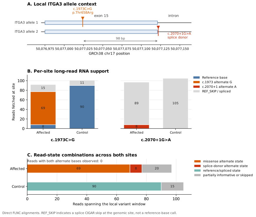
Figure 1. Long-read Iso-Seq RNA support for biallelic ITGA3 variants. **PacBio Iso-Seq direct FLNC alignments from hair follicle RNA were examined across the local ITGA3 region containing c.1973C>G, p.Thr658Arg and c.2070+1G>A. A, Schematic of the local ITGA3 allele context, showing the missense variant within exon 15 and the canonical splice-donor variant immediately downstream of exon 15. B, Per-site long-read RNA support showed alternate G reads at c.1973C>G and alternate A reads at c.2070+1G>A in the affected sample, whereas alternate-supporting reads were not observed in the unaffected control. C, Joint read-state analysis across both sites showed that the two alternate alleles were observed on separate RNA molecules, with no reads carrying both alternate bases. These data provide transcript-level support for expression of both ITGA3 variant alleles and are consistent with a compound heterozygous configuration. REF_SKIP indicates a splice-alignment skip over the genomic coordinate and should not be interpreted as a reference-base call.
Exploratory plasma proteomics nominates epithelial repair, cytoskeletal, and trafficking pathways
To investigate systemic proteomic alterations, we profiled patient plasma using the Olink Explore HT platform. Due to the single-patient study design, we compared the patient's proteomic profile against two healthy-donor reference pools (unstimulated and ex vivo stimulated) rather than using an inferential differential-abundance model. Protein levels—reported as principal component (PC)-normalized NPX values on a log2 scale—were evaluated using a NPX approach (NPX = patient NPX − healthy pool NPX). We focused on "concordant proteins," defined strictly as those passing assay quality control (QC) and exhibiting a uniform directional shift with an absolute NPX 2 across both healthy-donor contrasts.
Out of 5,401 QC-passing assays, 571 proteins met this stringency threshold, with 484 demonstrating concordant decreases and 87 showing concordant increases in the patient. The predominant systemic signature was a marked downregulation of proteins governing cilium assembly, membrane trafficking, and Rho-GTPase/cytoskeletal dynamics. This included prominent depletions in ciliogenesis and centrosomal factors (e.g., IFT88, IFT74, IFT20, OFD1, CEP135, CEP152, CEP250, TUBB8B, PKD2, RP2, CCP110), as well as trafficking and cytoskeletal regulators (e.g., DCTN1, DCTN2, RAB11FIP3, EXOC7, TRIP10, ARHGAP24, VAV3, WAS, WASF3, ARHGEF2, IQGAP2, ARF6, STX4, GGA1, GGA3, and BICD2). Over-representation analysis, performed using GSEApy9 against the MSigDB10 v2025.1 C2 canonical and Hallmark gene sets, nominated cilium assembly as the top down-concordant pathway, followed by membrane trafficking and Rho-GTPase signaling (Figure 2). Because no individual pathway reached a formal false discovery rate (FDR) < 0.05, we interpret these findings as a coherent, hypothesis-generating signal rather than definitive statistical proof of pathway enrichment.
Conversely, the smaller subset of 87 concordantly increased proteins was enriched for markers of epithelial repair and cellular stress biology, including TLE2, AKT2, HBEGF, AGR2, DDX60, CASP4, GAST, and RAD51. Collectively, these exploratory Olink results suggest that the patient's circulating proteome captures a state of disrupted cytoskeletal organization and mucociliary trafficking occurring alongside reactive epithelial repair. While this proteomic assay does not directly assess ITGA3 function, the observed systemic protein shifts are biologically congruent with impaired integrin-mediated epithelial adhesion and polarity, serving as a critical bridge between the genetic diagnosis and our subsequent tissue-level analyses.
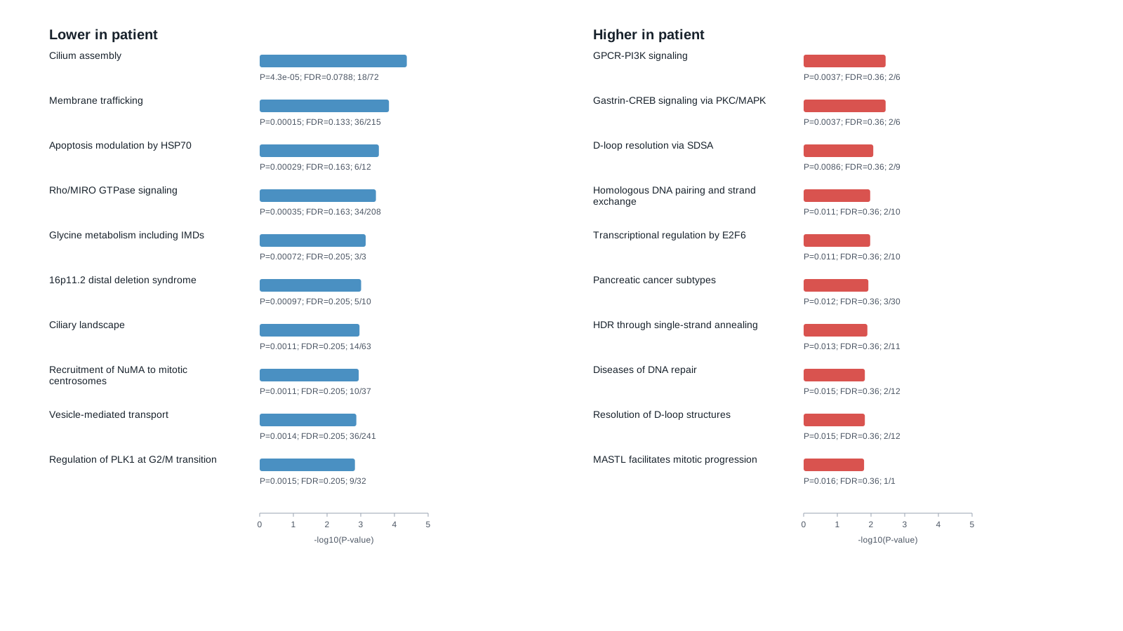
Figure 2. Olink pathway over-representation analysis.**The strongest protein-level pattern was a decrease in cilium assembly, membrane trafficking, and Rho-GTPase/cytoskeletal programs in the patient relative to both unstimulated and stimulated healthy-donor pools. These pathways are biologically consistent with impaired epithelial adhesion, polarity, and mucociliary trafficking in the setting of compound heterozygous ITGA3 variants, but they require orthogonal validation in airway tissue or patient-derived epithelial models.
PBMC single-cell RNA-seq suggests immune remodeling and inflammatory activation
To characterize the peripheral immune landscape, we performed single-cell RNA sequencing (scRNA-seq) on patient and control PBMCs. Following data integration, UMAP dimensionality reduction, and marker-guided manual annotation, we recovered 21,176 high-quality cells (10,078 patient; 11,098 control). We successfully resolved all major circulating immune lineages, including B cells, CD4 T cells, naive and effector/memory CD8 T cells, classical and non-classical monocytes, natural killer (NK) cells, CD56hi NK cells, myeloid dendritic cells, regulatory T cells (Tregs), platelets, and gamma-delta T cells (Figure 3A).
Comparative compositional analysis revealed marked shifts in the patient's peripheral immune balance (Figure 3C). The patient exhibited proportional expansions in CD4 T cells, classical monocytes, and naive CD8 T cells. Conversely, there was a relative depletion of effector/memory CD8 T cells, gamma-delta T cells, NK cells, B cells, and platelets. This compositional skewing aligns with chronic disease-associated immune remodeling11–12, characterized by the relative expansion of monocytic and CD4 compartments at the expense of cytotoxic and innate-like lymphocyte populations.
To identify the transcriptional programs underlying these shifts, we performed cell-type-resolved differential expression analysis using the Wilcoxon rank-sum test via the Scanpy rank_genes_groups function13 (Figure 3B). Comparisons were conducted both globally across all PBMCs and within specific populations containing at least 200 cells per group. A recurrent signal emerged surrounding antigen presentation: in the global all-cell comparison, HLA-DRB5 and HLA-DQA2 were downregulated in the patient, and the broader set of suppressed genes showed enrichment for antigen processing, presentation, and NK-cell-mediated cytotoxicity. This major histocompatibility complex (MHC) class II-related suppression was most pronounced in cytotoxic compartments (NK cells and effector/memory CD8 T cells). In contrast, several class II genes were upregulated within classical monocytes, indicating a highly cell-type-dependent reorganization of the antigen-presentation machinery.
Over-representation analysis of patient-upregulated genes against the MSigDB v2025.1 Hallmark and C2 canonical collections further supported an activated systemic inflammatory state (Figure 3D). The patient-enriched transcriptome was heavily skewed toward immune-activation programs, capturing robust signatures for allograft rejection, IL2-STAT5 signaling, TNF-alpha signaling via NF-kB, and broad inflammatory responses. Additional upregulated programs included cytokine-cytokine receptor interactions, hematopoietic cell lineage pathways, and FOXP3-related immune modules. Taken together, these scRNA-seq data uncover an inflammatory peripheral immune state marked by diminished cytotoxic lymphocyte pools, altered antigen presentation, and pronounced cytokine-driven immune activation. Given the single-case design, these findings represent exploratory systemic signatures that necessitate future validation in larger cohorts or orthogonal functional assays.
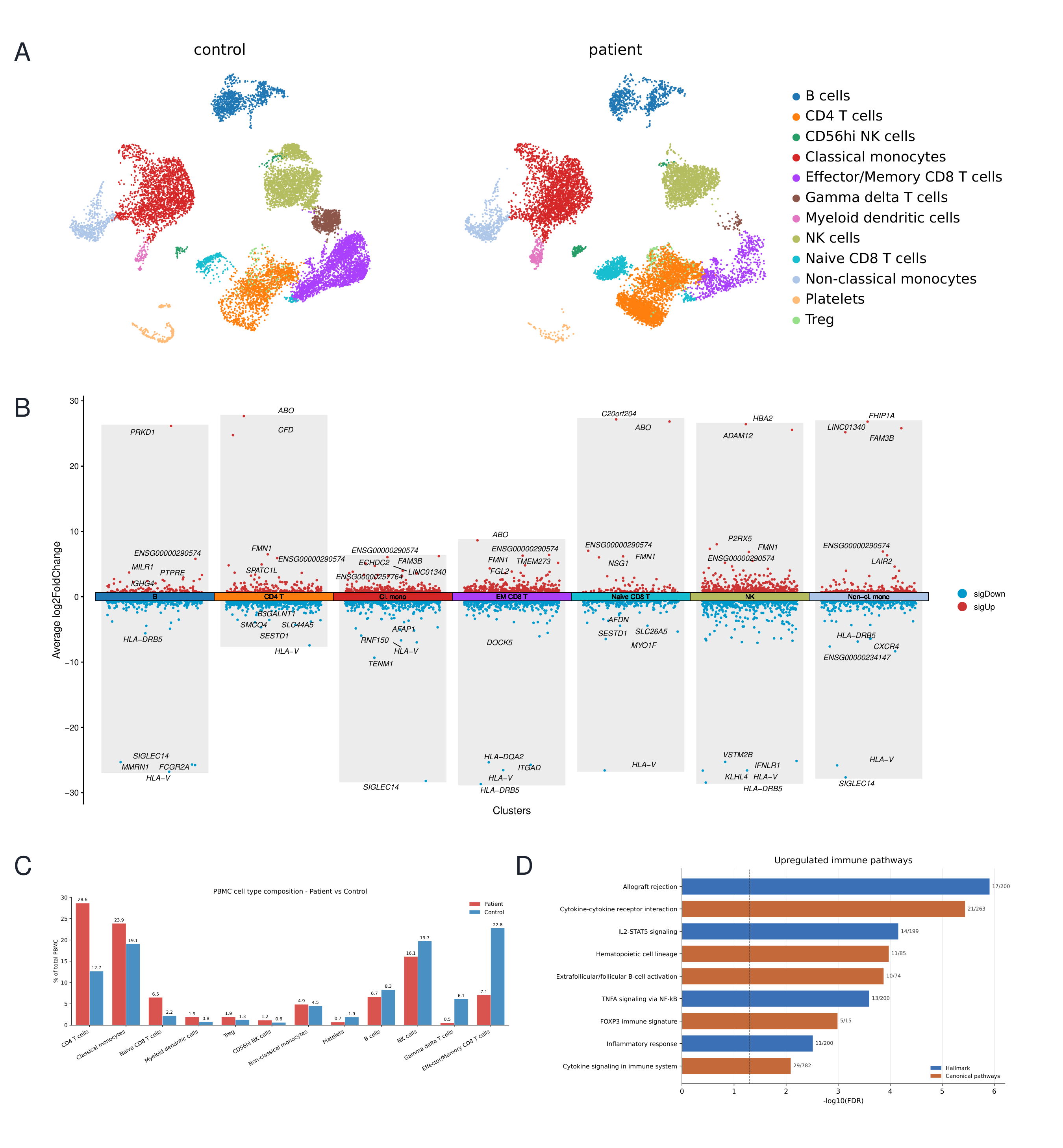
Figure 3. PBMC scRNA-seq results of patient-control immune remodeling.**A, UMAP of annotated PBMC populations from the patient and control samples. B, Cell-type-resolved differential-expression volcano plot for patient-versus-control comparisons; positive log2 fold change indicates higher expression in the patient. C, Relative abundance of annotated PBMC populations in patient and control samples. D, Over-representation analysis of patient-upregulated genes using MSigDB Hallmark and C2 canonical pathway collections.
Lung spatial transcriptomics identifies a fibrotic DATP-like transitional AT2-fibroblast remodeling niche
To dissect the tissue architecture of progressive lung remodeling, we applied Xenium spatial transcriptomics to the patient's 2017 and 2022 lung biopsy sections. Following marker-guided annotation and refinement of the AT1 compartment, the integrated dataset resolved 1,332,326 cells across 14 distinct epithelial, stromal, endothelial, and immune populations (Figure 4A). Fibroblasts constituted the largest cellular compartment at both timepoints, expanding slightly from 28.81% in 2017 to 30.04% in 2022 (Figure 4B). Notably, transitional AT2 cells underwent a marked longitudinal expansion, doubling from 12,425 cells (2.01%) in 2017 to 25,157 cells (3.52%) in 2022. This pronounced increase nominated the transitional AT2 compartment as a primary epithelial driver of progressive fibrotic remodeling.
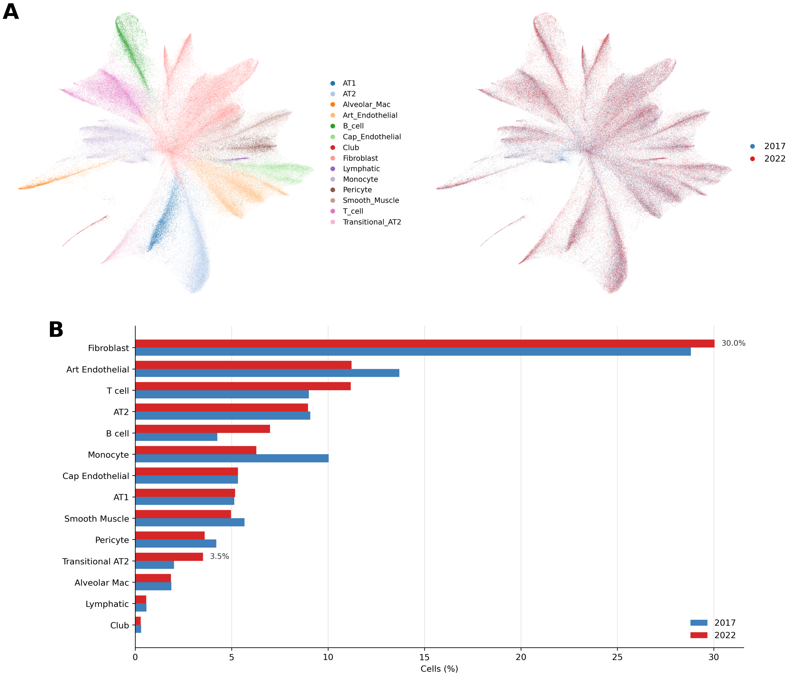
Figure 4. Spatial profiling captures progressive longitudinal expansion of transitional AT2 cells.** (A) UMAP of Xenium spatial transcriptomics annotations, resolving 14 distinct cell populations across the 2017 and 2022 lung biopsies. (B) Relative cellular abundance demonstrating the stability of the dominant fibroblast compartment and the marked longitudinal expansion of the transitional AT2 population.
Pathway scoring further solidified the transitional AT2 population as a central hub of disease progression. We evaluated a panel of predefined lung remodeling axes—including ITGA3, basement membrane, epithelial injury, AT2 stress, aberrant basaloid, senescence/SASP, fibrotic fibroblast, SPP1 macrophage, TGF-β/YAP/WNT, and ECM remodeling modules14–16 (Figure 5B)—and mapped the transcriptomic shifts across refined AT1-canonical lineages from 2017 to 2022 (Figure 5A). Transitional AT2 cells exhibited substantial longitudinal score increases in epithelial injury (+0.194), aberrant basaloid (+0.201), senescence/SASP (+0.105), TGF-β/YAP/WNT (+0.114), ECM remodeling (+0.125), and fibrotic-fibroblast (+0.056) modules. Spatial projections revealed recurrent, heterogeneous clustering of these signatures across the tissue (Figure 5C, D). Conversely, the SPP1 macrophage score peaked exclusively within alveolar macrophages, indicating a parallel but distinct immune remodeling process. Together, these spatial mappings indicate that disease progression is not characterized by a global, pan-tissue upregulation of stress pathways, but rather by the targeted amplification of specific epithelial and injury programs strictly confined within their respective cellular compartments.
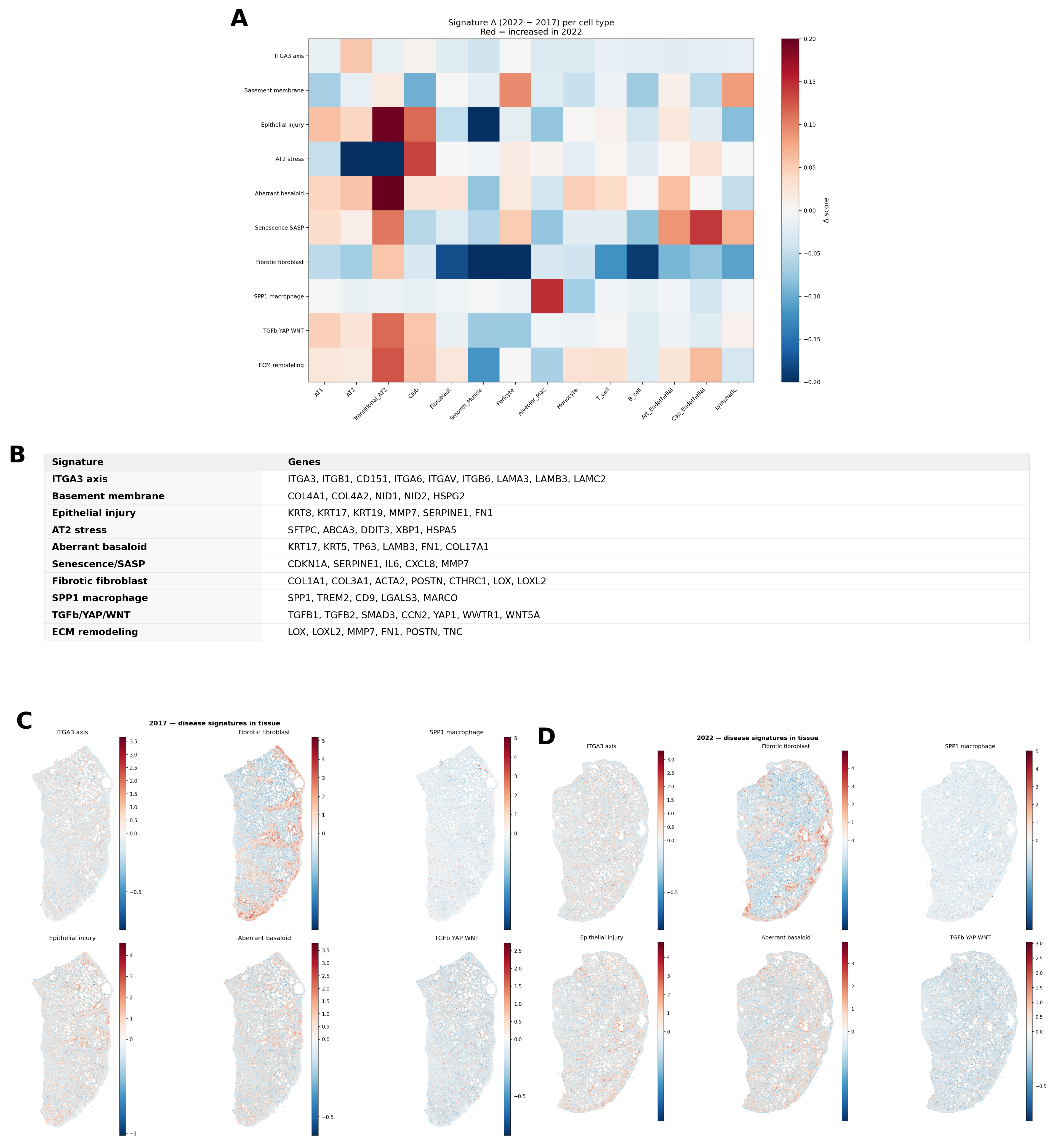
Figure 5. Spatial and longitudinal distribution of specific fibrosis-axis signatures.** (A) Heatmap quantifying longitudinal shifts (2017 to 2022) in predefined fibrosis and remodeling module scores across refined AT1-canonical lineages. (B) Gene sets comprising the evaluated fibrosis-axis signatures. (C, D) Spatial coordinate mapping of representative disease-signature scores across the (C) 2017 and (D) 2022 lung tissue sections.
Given their prominent variation across multiple stress axes and their canonical role as progenitors for AT1 replacement, we hypothesized that stalled transitional AT2 cells actively orchestrate aberrant remodeling. Spatial mapping placed both canonical AT2 and transitional AT2 cells squarely within remodeled, fibroblast-dense regions (Figure 6A, B). Fibroblast-anchored Squidpy co-occurrence analysis17 quantitatively confirmed this enrichment, revealing a strong spatial relationship that intensified by 2022 (Figure 6C). Specifically, the transitional AT2-to-fibroblast peak co-occurrence ratio increased from 1.194 (at ~89 µm) in 2017 to 1.321 (at ~113 µm) in 2022, reaching a maximum differential of +0.139 at ~125 µm. The strengthening of this intermediate-distance spatial correlation underscores the active consolidation of a shared epithelial-fibroblast remodeling niche as the disease advances.
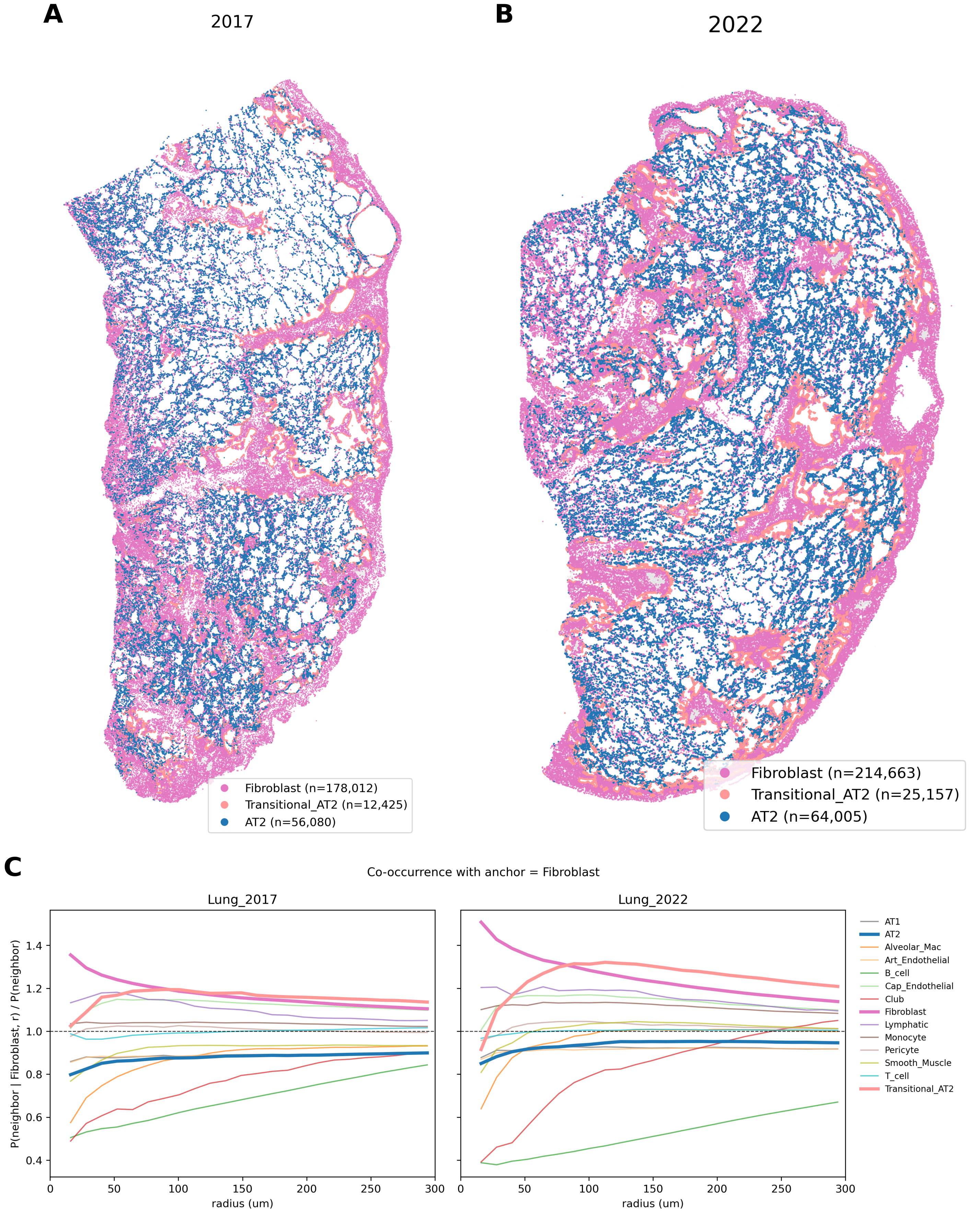
Figure 6. Spatial co-localization defines an active transitional AT2-fibroblast niche.** (A, B) Tissue coordinate maps highlighting refined AT2, transitional AT2, and fibroblast populations in the (A) 2017 and (B) 2022 sections. (C) Fibroblast-anchored Squidpy spatial co-occurrence analysis demonstrating an intensifying intermediate-distance spatial correlation between transitional AT2 cells and fibroblasts over time.
To contextualize this spatial architecture, we benchmarked our patient data against an external pulmonary fibrosis cohort (Vannan et al.)18. Unsupervised niche-composition analysis demonstrated that the patient's macro-level spatial architecture remained relatively stable between 2017 and 2022, aligning closely with the complex niche composition of the Vannan disease cohort rather than healthy controls (Figure 7A). However, cross-dataset disease-signature scoring revealed a highly distinct molecular trajectory underlying this shared fibrotic architecture. Most notably, the patient exhibited a strong positive ITGA3-axis signal—a feature absent in the broader Vannan disease cohort. Furthermore, while the patient experienced localized longitudinal increases in classical fibrosis drivers, their absolute activation of Senescence-SASP and TGF-β/YAP/WNT pathways remained markedly lower than the Vannan cohort baseline. Despite this macro-level architectural stability, signature scoring captured aggressive pathway-level progression in the patient, particularly a sharply escalating aberrant-basaloid signature by 2022 (Figure 7B). This benchmarking confirms the patient's alignment with general fibrotic tissue states while highlighting a unique, ITGA3-specific molecular etiology distinct from classical idiopathic pulmonary fibrosis (IPF).
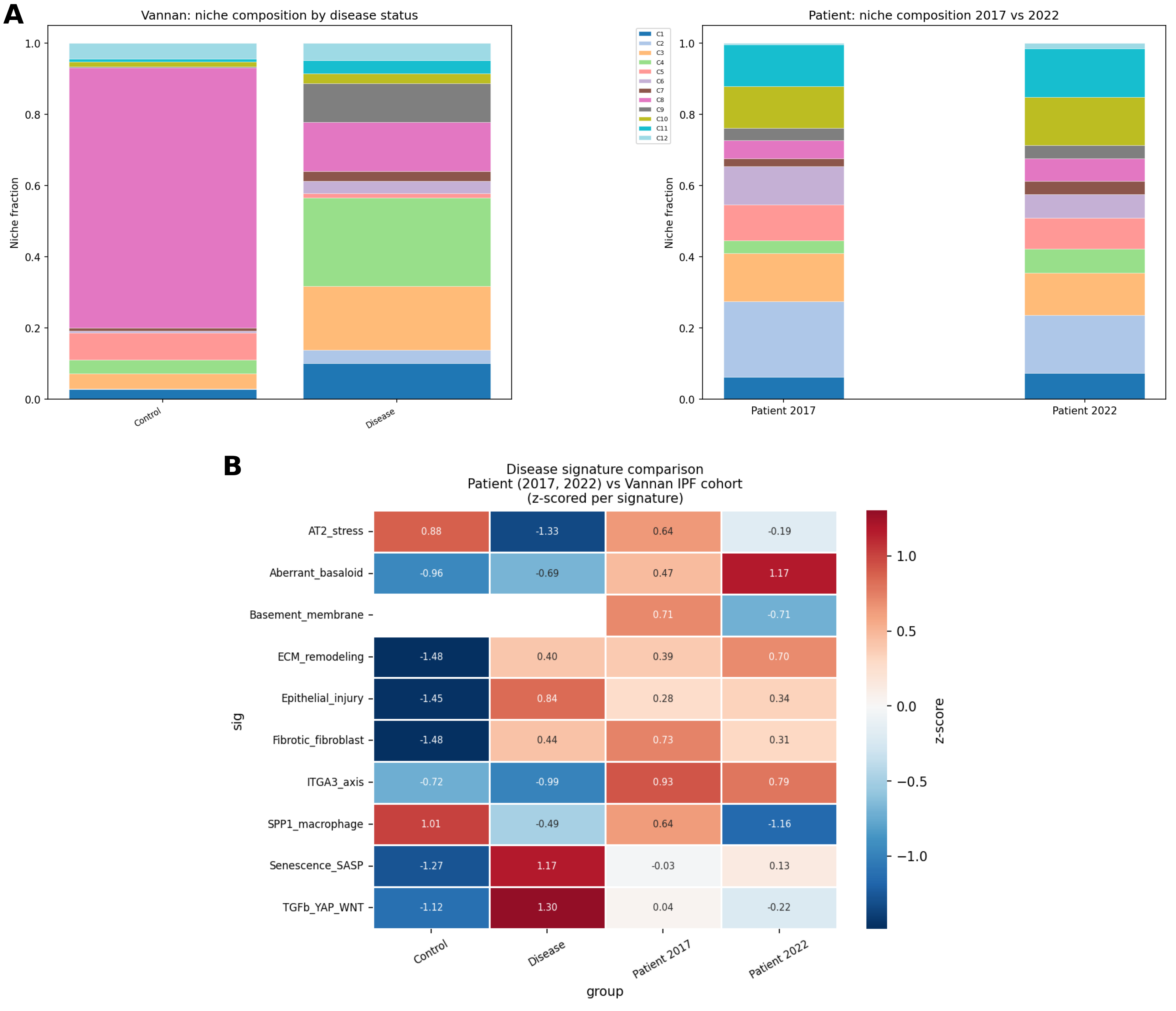
Figure 7. Molecular divergence of ITGA3-driven fibrosis from classical idiopathic pulmonary fibrosis (IPF).** (A) Unsupervised niche-composition benchmarking of the patient’s spatial architecture against the external Vannan et al. control and IPF disease cohort. (B) Cross-dataset disease-signature heatmap demonstrating a distinct molecular trajectory in the patient, characterized by high ITGA3-axis and aberrant-basaloid signatures, but lower baseline TGF-β/YAP/WNT activation compared to classical IPF.
To determine whether these transitional AT2 cells represent a physiological repair intermediate or a persistent damage-associated state, we mapped our data against a murine bleomycin-induced AT2-injury reference19. Cross-species delta-correlation analysis revealed that the patient’s transitional AT2 pseudobulks clustered distinctly with the mouse bleomycin-induced fibrotic damage-associated transient progenitor (DATP) state, rather than with healthy AT2 baselines or regenerative intermediates (Figure 8A). While absolute correlation scores were predictably modest due to cross-species panel divergence (Pearson r = 0.279, Spearman r = 0.424 across 4,747 shared genes), UMAP visualization supported localized patient-specific increases in profibrotic and aberrant-basaloid signatures (Figure 8B). Longitudinally, the generic DATP-core score increased from 2017 to 2022, but the most severe exacerbations occurred within the aberrant-basaloid and fibrotic-DATP modules. These cross-species findings strongly indicate that the patient’s transitional AT2 population represents a pathologically active, fibrotic epithelial injury state driving niche remodeling, rather than a transient physiological repair phase.
Crucially, this fibrotic DATP-like program connects our spatial observations to targetable stress-mediated fibrosis pathways. Pathologic activation of the ER-stress sensor IRE1α has been shown to force AT2 cells into a fibrotic DATP state—a transition driven by regulated IRE1-dependent decay (RIDD) rather than canonical XBP1 splicing19. Consistent with this model, the patient's transitional AT2 cells highly expressed associated DATP and aberrant-basaloid markers (KRT17, KRT8, CDKN1A, GDF15, and ITGB6), which were detected in 60.56% and 57.53% of these cells in 2017 and 2022, respectively. Because integrin beta-6 (ITGB6) directly mediates epithelial TGF-β activation, its robust expression suggests the patient's alveolar remodeling is actively driven by this IRE1α-fibrotic axis. Furthermore, the patient's mixed restrictive and obstructive clinical phenotype included severe constrictive bronchiolitis—a small-airway pathology distinct from alveolar remodeling. To investigate whether a shared stress mechanism was driving this parallel disease process, we evaluated IRE1α-axis markers across other epithelial compartments (Figure 8C). This ER-stress/unfolded protein response (UPR) signature was not restricted to the alveoli; club cells, which govern bronchiolar epithelial repair, exhibited a massive longitudinal exacerbation of these pathways. By 2022, club cells showed a dramatic upregulation of XBP1 (mean log1p 1.622 to 2.585; FDR < 1e-300) alongside a simultaneous surge in their DATP-core score (0.278 to 0.576; FDR = 6.69e-23). Collectively, these results suggest that IRE1α/UPR-related epithelial stress independently drives parallel pathologies in this patient—alveolar fibrosis via transitional AT2 cells and constrictive bronchiolitis via club cells—nominating the IRE1α axis as a unifying therapeutic target.
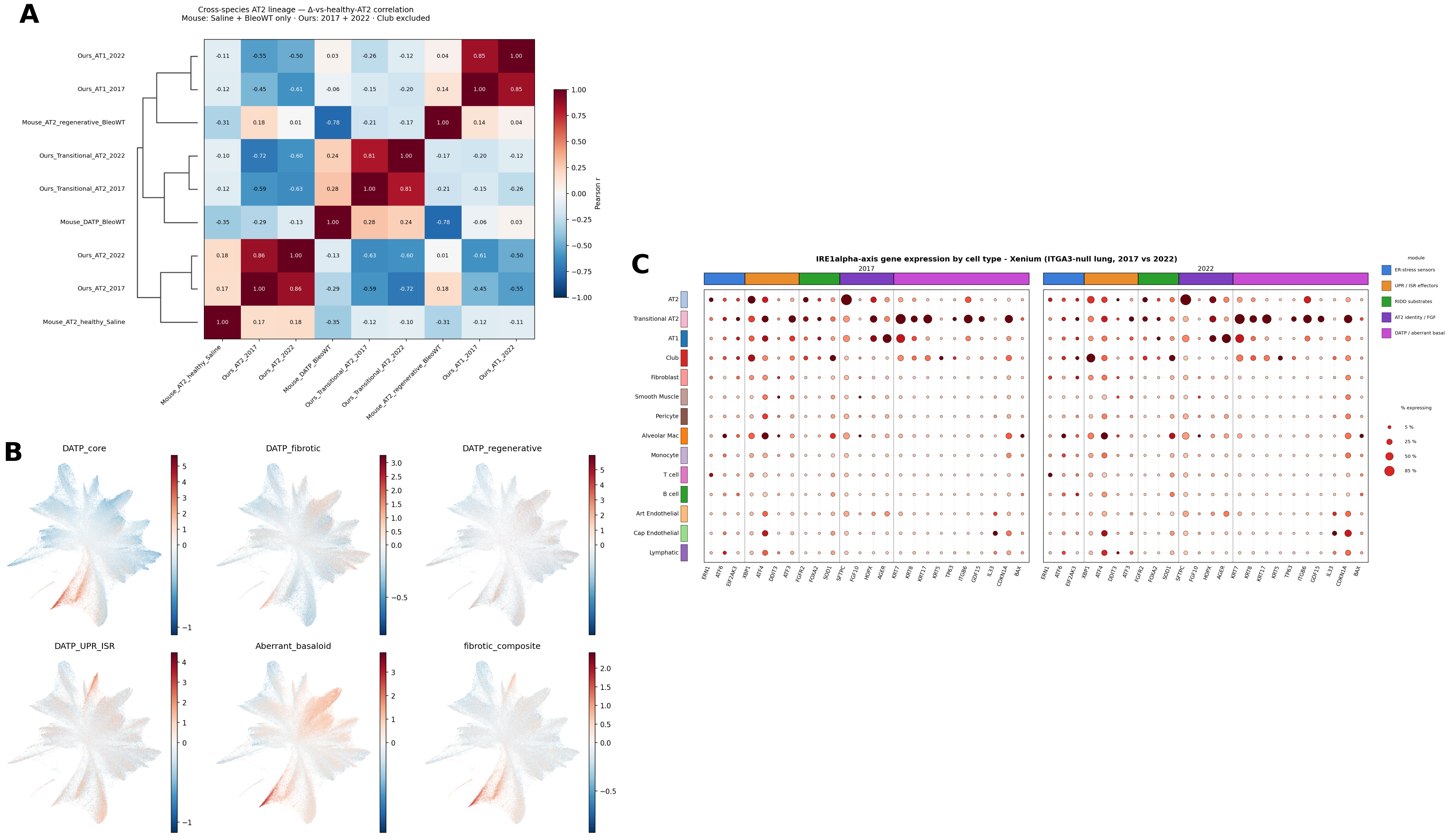
Figure 8. A shared IRE1α stress axis drives both fibrotic DATP-like AT2 cells and aberrant club cell remodeling. **(A) Cross-species delta-correlation heatmap mapping patient epithelial states against a murine bleomycin-induced AT2-injury reference, aligning patient transitional AT2s with the fibrotic DATP state. (B) UMAP projection of DATP-associated signature scores. (C) Dot plot detailing the longitudinal exacerbation of ER-stress sensors, UPR effectors, and DATP/aberrant-basal markers across both alveolar (AT2) and small-airway (club cell) compartments, highlighting a shared IRE1α-driven stress response driving parallel interstitial and bronchiolar pathologies.
Discussion
Pathogenic biallelic variants in ITGA3 cause severe multisystem disease, but the tissue programs linking impaired epithelial adhesion to progressive lung remodeling remain elusive. By integrating genetic prioritization, long-read transcriptomics, systemic proteomics, and longitudinal spatial profiling, we mapped the patient-specific tissue architecture of ITGA3-associated disease. Our spatial data revealed that progressive fibrosis is driven by an expanding, damage-associated transient progenitor (DATP)-like transitional AT2 population that progressively anchors to fibroblasts, forming an active remodeling niche. Crucially, we identified a shared IRE1α/UPR stress axis driving parallel pathologies: the fibrotic DATP-like state in the alveoli and aberrant club cell remodeling in the small airways, directly correlating with the patient's mixed restrictive-obstructive clinical phenotype.
Clinically, this case highlights how integrating transcriptomic and spatial data with genomic sequencing can transform a static molecular diagnosis into actionable biological insight. Long-read RNA sequencing confirmed the active trans expression of both ITGA3 variants, while spatial mapping pinpointed the resulting localized epithelial stress. Therapeutically, the pervasive IRE1α/UPR-driven stress signatures suggest that conventional, broad-spectrum immunosuppression may be insufficient. Instead, interventions targeting fibrotic epithelial states, localized TGF-β activation, or ER-stress pathways represent rational mechanistic avenues for treating ITGA3-associated fibrotic lung disease.
We acknowledge the inherent limitations of an exploratory, single-patient study. Spatial transcriptomics cannot definitively uncouple disease progression from regional tissue heterogeneity or treatment effects, and our proposed DATP-like and IRE1α-driven mechanisms require orthogonal functional validation in patient-derived in vitro models. Nevertheless, in rare Mendelian disorders where large cohorts are unfeasible, this deep multi-omic phenotyping effectively bridges the gap between molecular diagnosis and tissue-level pathophysiology, prioritizing the IRE1α/UPR stress axis as a central mechanism and candidate therapeutic target.
Conclusion
In conclusion, this study uses an unusually broad set of patient-derived assays to connect biallelic ITGA3-associated disease with systemic immune and proteomic remodeling and, most importantly, spatially resolved fibrotic lung architecture. The lung spatial data nominate DATP-like transitional AT2 cells, fibroblast-associated remodeling neighborhoods, and epithelial stress programs as candidate mechanisms linking impaired epithelial adhesion to progressive fibrosis and small-airway disease. Although these findings are limited by the single-patient design and require functional validation, they illustrate how integrated multi-omics can move rare-disease analysis from molecular diagnosis toward patient-specific mechanistic hypotheses and precision-medicine reasoning.
Be the first to comment on this publication.